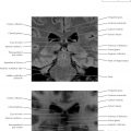
Brain Axial 1
Normal Anatomy
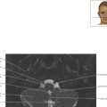
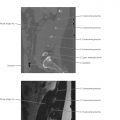
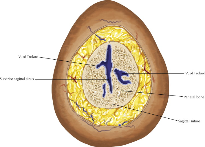
Named after the French anatomist Paulin Trolard (1842–1910) and also known as the superior anastomotic vein, the vein of Trolard is the largest cortical vein at the convexity (see also Brain Coronal 15 ). The vein of Trolard anastomoses with the middle cerebral vein and the superior sagittal sinus.
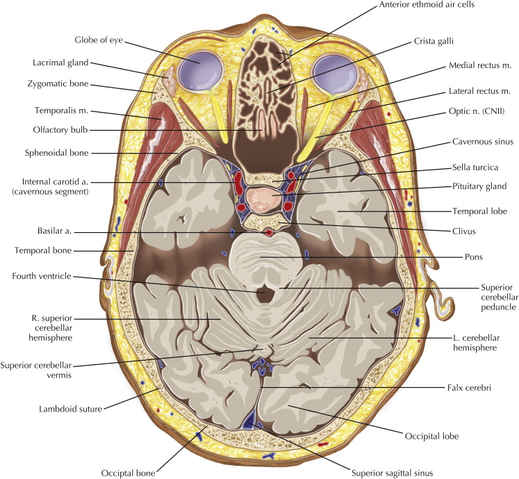
Note the first cranial nerves, the olfactory nerves, in the olfactory grooves along the medial inferior floor of the anterior cranial fossa. Please note there is volume averaging of cortical veins with the top of the calvaria such that the veins artifactually appear to be within the bone rather than within the cranial vault (see Brain Coronal 2 ).
Diagnostic Consideration
A lesion, such as a metastatic lesion in the marrow, can often be missed at the vertex (vertex cranii). Diffusion-weighted magnetic resonance imaging (MRI) is often useful in identifying such lesions, not because the lesion is diffusion restricted, but because all the other structures are often low in signal, making the lesion conspicuously bright. The same principle holds for the base of the skull, where the presence of many structures can make pathologic lesions easy to overlook.


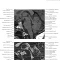
Brain Axial 5

Normal Anatomy
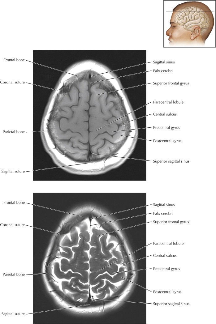
The “omega sign” denotes a sulcal configuration similar to the Greek letter Ω and is one sign of the central sulcus separating the motor strip anteriorly and the sensory cortex posteriorly. The omega sign also demarcates the frontal lobe from the parietal lobe. Note that the two sulci in the midline immediately posterior to the medial ends of the central sulci should resemble a smile.


Brain Axial 9

Normal Anatomy
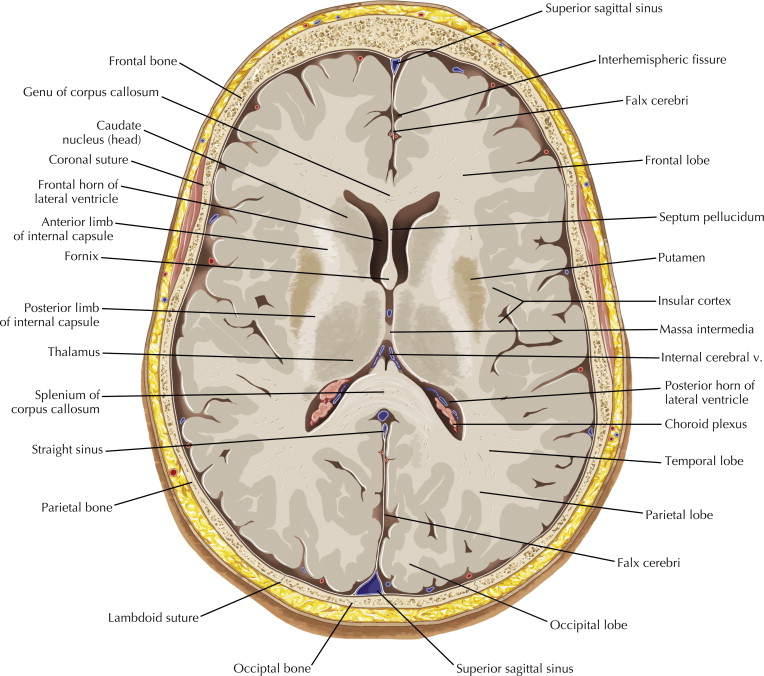
The white matter fibers at the level of the basal ganglia known as internal capsule ( Axials 10 and 11 ) are known as corona radiata ( Axials 8 and 9 ) more superiorly along the lateral margins of the ventricles, and as the centrum semiovale superior to the ventricles ( Axial 7 ).

Brain Axial 10
Normal Anatomy
Note how the signal of the basal ganglia is not ideal on this MR sequence tailored to capture the entire brain. For better delineation, see Chapter 3 .

Brain Axial 11

Pathologic Process
The foramen of Monro (interventricular foramen) can be an important point of obstruction of cerebrospinal fluid (CSF) flow. A small colloid cyst can occasionally be found at this location and could cause obstruction in a relatively short time, rapidly becoming fatal if untreated. CSF is produced at a rate of about 500 mL/day and the subarachnoid space around the brain and spinal cord can contain only 150 mL. CSF intracranially and within the spinal canal turns over about four times a day.

Brain Axial 12

Normal Anatomy
The habenula (Latin habena, “rein”) originally referred to the pineal gland stalk but is now commonly used to indicate a nearby group of neurons in the dorsal and caudal thalamus. The insular cortex is often the first location where an infarct in the area of the middle cerebral artery will become evident on unenhanced computed tomography (CT) of the head. Loss of differentiation of the insular gray matter from the subinsular white matter should always be evaluated.


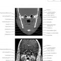
Brain Axial 14

Normal Anatomy
Note the small but conspicuous paired mammillary bodies anterior to the interpeduncular cistern, at the anterior end of the fornices. The mammillary bodies are part of the limbic system and are believed to add the element of smell to memories. The bodies can be enlarged and T2-weighted bright in Wernicke’s encephalopathy. Do not confuse the mammillary bodies with the paired inferior and superior colliculi, which are located on the posterior surface of the brainstem.

Brain Axial 15

Normal Anatomy
Note that the basal cisterns are composed of the suprasellar cistern, which resembles a six-pointed star, and the smile-shaped quadrgeminal plate cistern (see also Axial 14 ). The six points of the suprasellar cistern include the interhemispheric fissure, two Sylvian cisterns, two ambient cisterns, and the interpeduncular cistern.
Pathologic Process
The interpeduncular cistern is a good anatomic feature to scrutinize; this location often first reveals subarachnoid hemorrhage when a brain aneurysm ruptures. This usually occurs from the area of the circle of Willis, which is prone to many anatomic vascular variations.

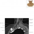
Brain Axial 16

Diagnostic Consideration
The temporal horns can be useful to help differentiate true hydrocephalus from ex vacuo dilation of the ventricles from central white matter volume loss. In the former case, the temporal horns are dilated and in the latter case they are not. The body of the lateral ventricles can sometimes look prominent but with normal-sized temporal horns where there is central white matter volume loss from chronic microangiopathic disease. Since the body and temporal horns are continuous structures, both should be dilated in true hydrocephalus.


Brain Axial 18