and Laurence Loeuillet2
(1)
Centre d’échographie Ambroise Paré, Les Mureaux, France
(2)
Hôpital Cochin, Paris, France
Brain biometrics is a vital part of the examination of the fetal brain. Many parameters can be measured by examining the brain. In addition to discussing the conventional measurement of biparietal diameter and head circumference, this chapter will address the biometrics of atrial diameter, cisterna magna, corpus callosum, brainstem, and cerebral vermis. It will also address the measurement of interorbital distance, anomalies of which may be indicative of fetal brain anomalies.
In order to be reproducible and relevant, biometric measurements require rigor in terms of the planes and sections used and compliance with certain quality criteria.
A number of tables are provided at the end of this book for purposes of consultation.
Biparietal Diameter (BPD) and Head Circumference (HC)
Solid quality criteria have been established for the measurement of biparietal diameter and head circumference (Salomon et al. 2006):
The measurement plane must be symmetrical.
The plane must pass through the thalami, the cavum of the septum pellucidum, and the posterior part, and the cerebellum must not be visible.
The image of the brain must make up more than half the ultrasound image.
The calipers must be correctly positioned.
Figure 4.1 illustrates these different requirements.

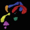
Fig. 4.1
Quality criteria for the measurement of BPD and HC: Green rectangle cavum of the septum pellucidum, pink triangle thalami, red circle cerebellum not visible, yellow line symmetry, turquoise circle calipers, BIP biparietal diameter
Regarding the calipers, their position depends on the methodology used to construct the standard plot off which the measurements are read. For instance, if Collège Français d’Echographie Foetale (CFEF) standard plots are used, the calipers should be placed in the middle of the skull’s proximal and distal bones. If the plots used are those prepared by Snijdjers and Nicolaides (1994), the calipers should be placed at the external interface of the skull. This remark is, of course, also valid when positioning the ellipse used to measure head circumference (Fig. 4.2).

Fig. 4.2
Positioning the ellipse when measuring HC
Cisterna Magna
An axial plane passing through the cavum of the septum pellucidum and the cerebellum is used to measure the cisterna magna. The actual measurement is made on the midline between the posterior surface of the cerebellar vermis and the internal surface of the occipital squama (Snijders and Nicolaides 1994; Steiger et al. 1995).

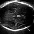
Fig. 4.3
Biometrics of the cisterna magna
Corpus Callosum
The corpus callosum may be measured for both its length and thickness (Achiron and Achiron 2001). Its length should be measured in a strict sagittal plane from the most anterior part of the genu to the most posterior part of the splenium. Its thickness should be measured in a median coronal plane bounded downward by the cavum of the septum pellucidum, upward by the echogenic line between the two hemispheres, and laterally by the frontal horns (Fig. 4.4).