and Laurence Loeuillet2
(1)
Centre d’échographie Ambroise Paré, Les Mureaux, France
(2)
Hôpital Cochin, Paris, France
The Comité National Technique de l’Echographie de Dépistage Prénatal (French National Committee on Prenatal Ultrasound Screening) indicated in its report published in 2005 that the ultrasound scan performed during the first trimester of pregnancy must check that the fetus does not present any cranial or midline anomalies. These recommendations aim to ensure that sonographers check that no acrania or holoprosencephaly is present. However, this first-trimester ultrasound scan may also be employed to screen for other anomalies of the cephalic extremity.
Acrania
The appearance of this anomaly will vary depending on the stage at which the fetus is examined. The brain will vary in appearance in relation to the extent to which brain tissue has disappeared. These malformations have been grouped together under the umbrella term “acrania,” and all, as common denominator, feature the absence of the bony dome of the skull.
It should be noted that amniotic fluid is often echogenic in acrania (Fig. 6.3b).
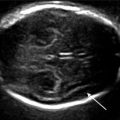
The discovery of acrania must prompt the sonographer to undertake a careful examination for amniotic band syndrome (Fig. 6.3c).
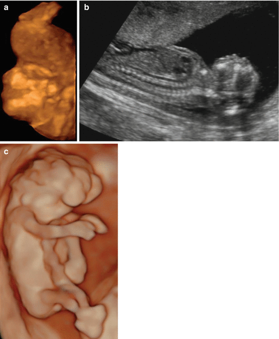


Figs. 6.1
(a, b) Acrania: (a) dome of the skull absent in maximum mode and (b) in sagittal image. (c) Exencephaly 10 gw + 6 days (3D ultrasound: HD live rendering method)
Fig. 6.2
(a, b) Acrania: images of exencephaly
Fig. 6.3
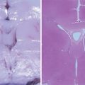
(a) Anencephaly: corresponding ultrasound image – pathology specimen. (b) Acrania: echogenic amniotic fluid. (c) Acrania: amniotic band syndrome (white arrow)
Holoprosencephaly
These midline anomalies share the common feature of showing merged midline structures. This may be more or less severe and produce a picture of holoproscencephaly ranging from the alobar form, where merger is almost total, to the lobar form, where only slight merger is seen. The alobar form is that most commonly detected in the first trimester, but semilobar forms may also be encountered at this point.
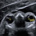
In alobar holoproscencephaly, the cerebral masses are completely merged (Fig. 6.4a), as are the lateral ventricles, giving the appearance of a single ventricle (Fig. 6.4b). These cephalic signs are often associated with facial anomalies such as the absence of nasal structures, marked hypotelorism or cyclopia, and possibly the presence of a proboscis (Fig. 6.4c).


Fig. 6.4
(a, b) Alobar holoproscencephaly: (a) cerebral masses merged, (b) single ventricle. (c) Alobar holoproscencephaly at 14 gw: anterior and posterior views of the brain, hypotelorism, and cleft (trisomy 13), fetal pathology specimens. (d) Proboscis. (e) Alobar holoproscencephaly: single ventricle (white arrow middle upper picture), cyclopia (white arrow middle picture median line) with two crystalline lenses, proboscis and absence of nasal structures, median cleft of palate (white arrow lower left picture). BIP Biparietal diameter
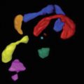
In the semilobar forms, the merger of the cerebral masses and lateral ventricles is incomplete, affecting only the anterior part of the brain.


Fig. 6.5
(a) Semilobar holoproscencephaly. (b) Semilobar holoproscencephaly at 15 gw, fetal pathology specimens: merger of the frontal lobes and arrrhinencephaly (a), single ventricular cavity to the front (c, d) separate hemispheres to the rear (b, e)
Opacities in Skull Images
Besides these major, conventional anomalies, the first-trimester ultrasound scan may also show skull anomalies: meningocele or meningoencephalocele (Figs. 6.6a, b), which are visible in the axial and sagittal planes. But as shown in Fig. 6.6a, special care is required to visualize meningoencephalocele.