and Laurence Loeuillet2
(1)
Centre d’échographie Ambroise Paré, Les Mureaux, France
(2)
Hôpital Cochin, Paris, France
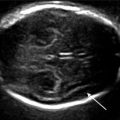
This brief review aims simply to describe the main steps in the development of the fetal brain and should allow ultrasonographers to familiarize themselves with the structures they will see during an early dating or first-trimester ultrasound.
The brain starts to form from the neural tube at the end of the 1st month of pregnancy. The appearance of folds and vesicles segments the neural tube into the forebrain or prosencephalon, midbrain or mesencephalon, and hindbrain or rhombencephalon.
Secondarily (around the 5th week of pregnancy), the prosencephalon itself subdivides into the telencephalon and the diencephalon, whereas the rhombencephalon divides into the metencephalon and the myelencephalon.
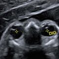
The optic stems and cups form from the diencephalon.
The cortex forms from the telencephalon, the cerebellum and the brainstem from the metencephalon, and the medulla oblongata from the myelencephalon.
Related posts:


Stay updated, free articles. Join our Telegram channel

Full access? Get Clinical Tree


