and Laurence Loeuillet2
(1)
Centre d’échographie Ambroise Paré, Les Mureaux, France
(2)
Hôpital Cochin, Paris, France
From 8 to 10 GW: Early Morphological Examination
From 8 to 10 gw
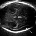
8 gw
 9 gw
9 gw
 10 gw
10 gw


Fig. 2.1
Corresponding histology from 8 to 10 gw: (a) 8 gw, (b) 9 gw, (c) 9 gw1/2, (d–f) 10 gw
Anatomical Landmarks
From 8 gw, the falx cerebri is visible (* telencephalic vesicles), and the rhombencephalic vesicle (°) is easily identifiable.
The cranial contours are visible from 9 gw.
The cerebral aqueduct becomes visible (arrow).
From 11 to 13 GW: Early Morphological Examination
11 gw


Fig. 2.2
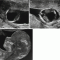
Corresponding histology at 11 gw. (a–c) Coronal sections, (d) axial section


Fig. 2.3
Corresponding histology at 12 gw. (a, b) Coronal sections, (c, d) axial sections


Fig. 2.4
Corresponding histology at 13 gw. (a, b) Coronal sections, (c) pathology specimen

Fig. 2.5
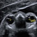
Fourth ventricle: histology at 13 gw (coronal sections) V4 intracerebral transluscency, CN cisterna magna
Anatomical Landmarks
The fourth ventricle (yellow arrows) is bordered by closure of the edges of the rhomboencephalic vesicles.
The third ventricle is clearly visible (turquoise arrows).
The upper part of the diencephalon (blue arrow) is visualized between the choroid plexuses of the lateral ventricles.
From week 12, the cerebral cortex can be visualized, and the orientation of Bichat’s fissures is clearly visible (dotted arrow). These fissures are oblique and form an acute angle with a posterior point. No Sylvian fissures are present (pink arrow).
From 14 to 16 GW: Morphological Examination


Fig. 2.6
16w: (a) Triplane, (b) Axial TIU, (c) Coronal TIU, (d) Sagittal TIU

Fig. 2.7
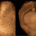
Corresponding specimens and histology at 15 gw
Anatomical Landmarks
14 gw
 14 gw
14 gw
 14 gw
14 gw

Anatomical Landmarks
At 14 gw, the cerebellar vermis (green arrow) can be seen behind the fourth ventricle (yellow arrows); the cisterna magna (yellow dotted arrow) can be distinguished behind the cerebellum.
The third ventricle (turquoise arrow) and the upper part of the diencephalon or roof of the third ventricle (blue arrow) are visible.
16 gw

16 gw

16 gw


Fig. 2.8
Corresponding specimens and histology at 16 gw: brain (a–d), cerebellum (e dorsal view), (f, g) Sagittal sections
Anatomical Landmarks
At 14 weeks, the fourth ventricle is clearly visible (yellow arrow).
The third ventricle (turquoise arrow) and the upper part of the diencephalon or roof of the third ventricle (blue arrow) are visible.
At 16 gw, the anterior part of the corpus callosum may be visible (orange arrow).
From 18 to 20 GW: Morphological Examination


Fig. 2.9
18 gw: (a) Triplane, (b) Axial TIU, (c) Coronal TIU, (d) Sagittal TIU

Fig. 2.10
Brain at 18 gw: (a) ventral view, (b) lateral view, (c) axial section, (d) sagittal section, (e) coronal section


Fig. 2.11
20 gw: (a) Triplane, (b) Axial TIU, (c) Coronal TIU, (d) Sagittal TIU
Anatomical Landmarks
18 gw

Fig. 2.12
Corresponding ultrasound images and fetal pathology specimens at 18 gw
18 gw

20 gw


Fig. 2.13
Fetal pathology specimen at 18 gw
Anatomical Landmarks
At 18 gw, the corpus callosum (red arrows) is already well developed and is visible in sagittal and coronal images. The Sylvian fissures (yellow arrows) are almost invisible.
At 20 gw, the corpus callosum is clearly visible and can be measured for thickness, length, and echogenicity (red arrows). The large commissures are recognizable by their upward scalloping and their marked echogenicity (green arrows). The Sylvian fissures are still discrete (yellow arrows) (see changes in Sylvian fissures).
20 gw


Fig. 2.14
Fetal pathology specimen at 20 gw: coronal section
20 gw


Fig. 2.15
Crura of fornix at 20 gw: (a) ultrasound images, (b) fetal pathology specimen
From 21 to 23 GW: Morphological Examination


Fig. 2.16
22 gw: (a) Triplane, (b) Axial TIU, (c) Coronal TIU, (d) Sagittal TIU

Fig. 2.17
Fetal pathology specimen: axial section at 21 gw