CHAPTER 15 Brain Stem
The brain stem is the narrow length of tissue that interconnects the spinal cord caudally with the brain rostrally.1–8 As such it extends from the transverse plane just caudal to the pyramidal decussation to the transverse plane just rostral to the superior colliculi.
Common terms and synonyms used throughout this discussion include the following:
Basis: the most ventral lamina of the full brain stem
Brain: the portion of the central nervous system rostral to the midbrain (i.e., the diencephalon and telencephalon)
Cerebral peduncles: Classically, the terms left and right cerebral peduncles signify all of the midbrain ventral to the cerebral aqueduct. On each side, a curved line drawn along the anterior surface of the substantia nigra subdivides the cerebral peduncle into a ventral crus cerebrorum and a dorsal tegmentum. As a consequence, the two crura cerebrorum form paired structures situated to each side of the interpeduncular fossa, while the tegmentum of the midbrain forms a single, bilaterally symmetric structure situated dorsal to the crura and ventral to the aqueduct. Presently, the term cerebral peduncle is most often used to indicate the crus cerebri.
Comb system: the bundles of striatal and pallidal fibers passing through the internal capsule
Corticobulbar: This term refers to fibers that descend from the cerebral cortex to any portion of the brain stem (not just the medulla). It derives from the old term bulb previously used to indicate the entire brain stem.
Crus (crura) cerebrorum: the most ventral portion of the midbrain ventral to the anterior border of the substantia nigra and lateral to the interpeduncular fossa
Lemniscal arc: This term is used to describe the group of ascending axons that, in aggregate, form the paired, transverse arches of white matter in the upper pons and midbrain. The structures of the lemniscal arc include the paired medial lemnisci medially, the paired lateral lemnisci laterally, and the paired spinothalamic tracts (spinal lemnisci) that angle over from lateral to medial as they ascend through the brain stem.
Sensory neurons: primary and secondary (relay). Each primary sensory neuron has (1) a peripheral process that extends to peripherally situated sensory endings, (2) a cell body that lies within the dorsal root ganglion or sensory ganglion related to a cranial nerve, and (3) a central process that enters the central nervous system via spinal or cranial nerves to synapse on second order neurons within the CNS. Secondary sensory (relay) neurons then receive the sensory data from the primary neuron and convey it onward to tertiary neurons and beyond.8
Tectum: the dorsal lamina of the midbrain situated dorsal to the aqueduct
Tegmentum: the middle lamina of the full brain stem situated between the basis ventrally and the aqueduct-fourth ventricle dorsally
Trapezoid body: the bundle of transverse fibers in the tegmentum of the pons that conveys auditory information bilaterally
ANATOMY
Longitudinal Divisions
From caudal to cranial, the brain stem is divided into three parts: the medulla oblongata inferiorly, the pons varolii in the middle, and the mesencephalon (midbrain) rostrally (Figs. 15-1 to 15-4). The caudal border of the medulla is a transverse plane situated just caudal to the decussation of the pyramids.4 The rostral border of the medulla and caudal border of the pons is the pontomedullary sulcus (ventrally) and the striae medullares at the floor of the fourth ventricle (dorsally). The rostral border of the pons and caudal border of the midbrain is the pontomesencephalic sulcus ventrally and a line drawn just inferior to the inferior colliculi dorsally. The rostral border of the mesencephalon and caudal border of the diencephalon is the line drawn along the medial mesencephalic sulcus and the inferior margins of the optic chiasm and tract (ventrally) and the posterior commissure (dorsally).4
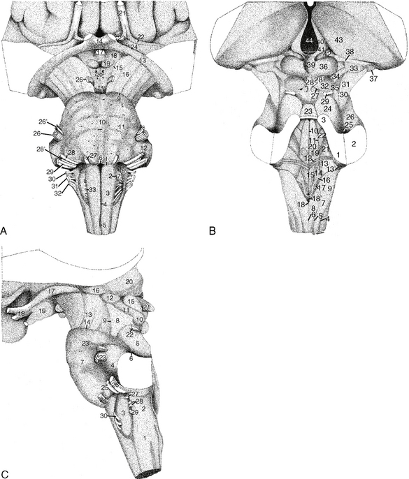
FIGURE 15-1 Surface of the brain stem and cranial nerves. Diagrammatic representations. A, Ventral view. B, Dorsal view, C, Lateral view with anterior to the reader’s left. A, Ventrally, the medulla is separated from the pons by the pontomedullary sulcus (1). The anterior median medullary sulcus (4) ascends between the paired medullary pyramids (3) to end superiorly at the foramen cecum (midline portion (6) of pontomedullary sulcus). Inferiorly, the anterior median sulcus shows a deflection (5) that indicates the site of the decussation of the pyramids. Laterally, the image displays the pyramids of the medulla (3), the preolivary sulcus (2) with the emerging rootlets of the hypoglossal nerve (CN XII), the olive (7), the supraolivary fossette (8), and the retro-olivary sulcus with the emerging rootlets of the glossopharyngeal, vagus and accessory nerves (CN IX, X, and medullary portion of XI). The pons is delimited inferiorly by the pontomedullary sulcus (1) and superiorly by the pontomesencephalic sulcus (9). The ventral aspect of the pons displays the median pontine sulcus (10), the transversely ridged “belly” of the pons (11), and the ventral aspect of the middle cerebellar peduncle (brachium pontis) (12). The abducens nerves (CN VI) (27) emerge from the pontomedullary sulcus 1 to 2 cm lateral to the midline. The facial and vestibulocochlear nerves (CN VII and VIII) (28 and 29) and the nervus intermedius (28′) emerge from the lateral aspect of the supraolivary fossette just inferior to the middle cerebellar peduncles. The smaller motor (26′) and larger sensory (26) roots of the trigeminal nerve emerge from the lateral aspect of the mid pons. The midbrain (mesencephalon) is delimited inferiorly by the pontomesencephalic sulcus (9) and superiorly by the inferior borders of the optic tract (13) and chiasm (14) and by the medial mesencephalic sulcus (15). The ventral aspect of the midbrain displays the crus cerebri (16) to each side of the interpeduncular fossa, and the oculomotor nerves (CN III) (25) emerging into the interpeduncular fossa from the medial aspects of each crus cerebri. The pituitary stalk (17), tuber cinereum (18), and mammillary bodies (19), of the hypothalamus of the diencephalon project inferiorly into the interpeduncular fossa but are not part of the brain stem. Laterally, the medial and lateral geniculate bodies project ventrolaterally from the thalamus (33 and 37 in B). B, Dorsally, the brain stem shows the rhomboid (diamond) shape of the fourth ventricle and the transversely oriented striae medullares (12) that subdivide that diamond into an inferior medullary triangle and a superior pontine triangle. The floor of the fourth ventricle shows the vertical median sulcus and the paired paramedian sulci limitantes (10 and 11) that flank the midline. The obex (18) lies at the inferior point of the fourth ventricle. The superior medullary velum forms the roof of the upper triangle. The lateral walls of the inferior fourth ventricle are formed by the inferior cerebellar peduncles (9). The lateral walls of the superior triangle are formed by the superior cerebellar peduncles (3). The middle cerebellar peduncles lie lateral to both the inferior and the superior cerebellar peduncles and do not contribute directly to the walls of the fourth ventricle. Further laterally, the groove (depression) between the superior and middle cerebellar peduncles is the parabrachial recess (25). Medulla. The medulla displays a “closed” portion situated inferior to the fourth ventricle and an “open” portion underlying the lower, medullary triangle of the fourth ventricle. The dorsal surface of the closed medulla shows the posterior median medullary sulcus (6), the paired posterior intermediate medullary sulci (5) and the posterolateral medullary sulci (4). The fasciculus gracilis ascends to the nucleus gracilis (8) just to each side of the median sulcus. The dorsal bulge caused by the gracile nucleus is designated the clava. The fasciculus cuneatus ascends to the nucleus cuneatus (7) between the posterior intermediate and posterolateral medullary sulci just lateral to the fasciculus and nucleus gracilis on each side. The dorsal surface of the rostral open medulla displays the hypoglossal trigone (15) medially, the vagal trigone (14) more centrally, and the medullary vestibular area (13) laterally. Pons. The dorsal surface of the pons forms the upper triangle of the floor of the fourth ventricle. Medially, it displays the median eminence (20) and the facial colliculus (19) that marks the site where the fibers of the facial nerve (CN VII) curve dorsal to the nucleus for the abducens nerve (CN VI). Superolaterally the trigeminal fovea (superior fovea) (22) marks the site of the underlying principal sensory and motor nuclei of the trigeminal nerve. The pontine vestibular area (21) lies far laterally at the lateral angle of the diamond. The dorsal cochlear nucleus (13′) forms the acoustic tubercle (13′) at the dorsal aspect of the inferior cerebellar peduncle. Midbrain. The superior (36) and inferior (32) colliculi take the shape of a butterfly with wider upper than lower wings. The cruciate sulcus is formed by a vertical arm that runs in the midline between the left and right colliculi and a horizontal arm that runs transversely between the superior and the inferior colliculi. The infracollicular sulcus delimits the inferior borders of the inferior colliculi. Inferior to the inferior colliculi, the frenulum veli acts as a midline suspensory ligament to elevate the superior medullary velum (23) over the fourth ventricle. Laterally, on each side, the brachium (34) of the inferior colliculus ascends to the medial geniculate body (33). The brachium (38) of the superior colliculus ascends over the medial geniculate body to connect with the optic tract, bypassing the lateral geniculate body (37). The lateral mesencephalic sulcus (30) delimits the dorsal edge of the crus cerebri (31). The trochlear nerves (CN IV) (29) emerge from the dorsal surface of the midbrain just inferior to the inferior colliculi, to each side of the frenulum veli. The pretectum (42), the pineal gland, the habenular trigones (41), and the striae medullares thalami (40) lie superior to the superior colliculi. C, Laterally, the medulla displays the lateral aspect of the pyramid ventrally, the hypoglossal rootlets (30) emerging from the pre-olivary sulcus, the olive (3), the rootlets of CN IX, X, and medullary XI (27, 28, and 29) emerging from the retro-olivary sulcus, the inferior cerebellar peduncle (2), and the lateral medullary funiculus (1). The rootlets of the abducens nerve (CN VI) (24), the facial nerve (CN VII) (25) and the vestibulocochlear nerve (CN VIII) (26) emerge along the pontomedullary sulcus in a line from paramedian (CN VI) into the supraolivary fossette (CN VII and VIII). The pons displays the profile of the belly of the pons, the rootlets of the motor (23′) and sensory (23) fibers of CN V, the middle cerebellar peduncle (4), the parabrachial recess (6), and the superior cerebellar peduncle (5). The midbrain displays the lateral aspect of the crus cerebri (13), the lateral mesencephalic sulcus (9), the lateral aspect of the midbrain (8) behind the crus, the superior cerebellar peduncle (5), the lateral aspects of the inferior colliculus (10) and its brachium (11), the superior colliculus (15), and the rootlets of CN IV (22) passing from the infracollicular groove around the lateral aspect of the mesencephalon. The optic chiasm (18), optic tract (17), lateral geniculate body (16), medial geniculate body (12), pineal gland (21), and thalamus (20) overhang the midbrain.
(Modified from Naidich TP, Duvernoy HM, Delman BN, et al. Duvernoy’s Atlas of the Human Brain Stem and Cerebellum: High-Field MRI, Surface Anatomy, Internal Structure, Vascularization and 3D Sectional Anatomy. New York, Springer, 2008.)
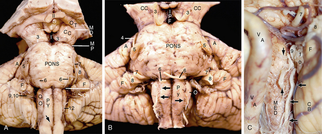
FIGURE 15-2 Surface of the brain stem and cranial nerves. Three specimens. A and B, Ventral surfaces of two gross specimens after removal of the arachnoid and blood vessels. Ventrally, the mesodiencephalic junction (MD) is the lower margin of the optic chiasm and optic tract. The mesopontine junction (MP) is the curved pontomesencephalic sulcus. The pontomedullary junction (PM) is the curved pontomedullary sulcus. Midbrain. The midbrain gives rise to two cranial nerves. CN III (3) exits the brain surface at the medial aspect of the crus cerebri (CC) and traverses the interpeduncular fossa (I, IPF) en route to the lateral dural wall of the cavernous sinus. CN IV (4) exits the brain surface contralateral to its nucleus of origin, emerges just inferior to the inferior colliculus (see Fig. 15-3B), and courses around the midbrain to enter the lateral dural wall of the cavernous sinus immediately inferior to CN III. In this specimen, the cut anterior end of CN IV has fallen over the upper pons and trigeminal nerve (5). m, paired mammillary bodies. Pons. The pons gives rise to four cranial nerves. CN V (5) crosses the dorsal brain surface at the upper lateral pons just anterior, inferior and medial to the overhanging anterior angle (A) of the cerebellum. CN VI (6) exits the brain surface at the pontomedullary sulcus, 1 to 2 cm lateral to the midline, courses anteriorly, superiorly, and slightly laterally to exit the skull via Dorello’s canal through the clivus. The arrow indicates the decussation of the pyramids. In B, the cut ends of CN VI have fallen to overlie the pontomedullary sulcus. CN VII (7) and CN VIII (8) cross the brain surface within the supraolivary fossette just superior to the upper pole of the inferior olive (O), just inferior to the middle cerebellar peduncle (MCP), and just ventromedial to the flocculus (F). These nerves exit the skull through the internal auditory canals. Medulla. The medulla gives rise to four cranial nerves. Cranial nerve IX (9) and cranial nerve X (10) cross the brain surface at the retro-olivary sulcus, dorsal to the olive (O), and traverse the jugular fossa to their distal innervations. CN XI (not labeled) crosses the brain surface at the medulla (medullary root) and at the lateral surface of the cervical spinal cord (spinal accessory root) and traverses the jugular fossa to its distal innervations. The rootlets (12, black arrows in B) of cranial nerve XII cross the brain surface at the preolivary sulcus, ventral to the olive (O), and exit the skull base via the hypoglossal canal. C, Coned-down lateral view. From ventral to dorsal, the vertebral artery (VA), medulla (MED), and the cerebellum (CB) form parallel vertical columns. The spinal accessory nerve (CN XI) (horizontal black arrows) ascends along the lateral surface of the medulla caudal to, and in line with, the rootlets of the glossopharyngeal (9) and vagal (10) nerves. The rootlets of the hypoglossal nerve (vertical black arrows) lie ventrally. The intervening olive is largely obscured by the nerve roots. As in A, the facial (CN VII) (7) and vestibulocochlear (CN VIII) (8) nerves emerge from the supraolivary fossette just below the middle cerebellar peduncle and just superior and ventral to the flocculus (F).
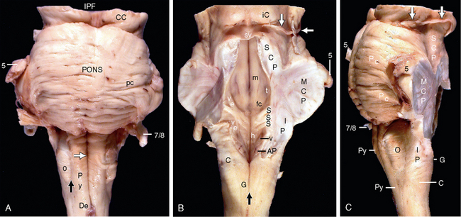
FIGURE 15-3 Gross anatomy of the brain stem. Cleaned ventral (A), dorsal (B) and lateral (C) surfaces of the medulla, pons, and inferior portion of midbrain, after removal of the arachnoid and blood vessels. The cerebellum was resected by sectioning the inferior (IP), middle (MCP), and superior (SCP) cerebellar peduncles. A, The ventral surface displays the anterior median medullary sulcus (white arrow), the paired medullary pyramids (Py) that flank the sulcus, the decussation (De) of the pyramids, the preolivary sulcus (black arrow), the olive (O), and the transversely oriented pontocerebellar fibers (pc) that give the pons its corrugated appearance. The facial (CN VII) (7) and vestibulocochlear (CN III) (8) nerves arise from the lateral portion of the pontomedullary junction. The trigeminal nerves (CN V) (5) arise from the lateral surface of the upper pons. A very small segment of the upper midbrain is seen superiorly, including parts of the crura cerebrorum (CC) and the interpeduncular fossa (IPF). B, The dorsal surface displays the rhomboid (diamond) shape of the fourth ventricle, the prominent midline median sulcus, and the transversely oriented striae medullares (S, S, S). The striae divide the floor of the fourth ventricle into a lower medullary triangle situated inferior to the striae and an upper pontine triangle situated superior to the striae. The lateral walls of the lower fourth ventricle are formed by the inferior cerebellar peduncles (IP). The lateral walls of the upper fourth ventricle are formed by the superior cerebellar peduncles (SCP). The middle cerebellar peduncles (MCP) lie lateral to both the ICP and the SCP, so they do not form part of lateral walls of the fourth ventricle. The medulla displays two portions: a closed lower portion of the medulla situated inferior to the fourth ventricle and an open upper portion of the medulla situated ventral to the medullary triangle of the fourth ventricle. The posterior median medullary sulcus (vertical black arrow) and the flanking fasciculi and nuclei graciles (G) terminate at the lower margin of the fourth ventricle medially. The paired fasciculi and nuclei cuneati (C) lie lateral to the gracilis, so they must ascend higher to terminate at the lower lateral border of the fourth ventricle. The floor of the medullary triangle displays a median sulcus, paired inferomedial hypoglossal trigones (h) marking the hypoglossal nuclei, and paired superolateral vagal trigones (v) marking the dorsal vagal nuclei. The area postrema (AP) lies caudal to the vagal trigone. At the pontine triangle, the floor of the fourth ventricle displays the paired medullary eminences (m) to each side of the median sulcus, the prominent hillocks of the facial colliculi (fc), at the lower ends of the medullary eminences, and the slight depressions of the foveae trigeminales (t) that mark the motor and principal sensory trigeminal nuclei on each side. The lateral angles of the fourth ventricle are occupied by broad vestibular areas (ve) that extend through both the medullary and the pontine triangles. C, The lateral surface of the brain stem is oriented with the ventral side to the reader’s left. The medulla shows the pyramids (Py) ventrally, the olive (O) laterally, and the inferior cerebellar peduncle (IP), fasciculus cuneatus (C), and fasciculus gracilis (G) dorsally. Cranial nerves VII and VIII (7/8), V (5), and IV (white arrows in B and C), the middle cerebellar peduncle (MCP), and the superior cerebellar peduncle (SCP) are also labeled.
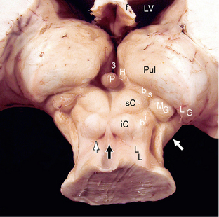
FIGURE 15-4 Posterior superior view of the midbrain and diencephalon. The quadrigeminal plate (tectum) of the midbrain displays the “butterfly” pattern of the paired superior colliculi (sC) extending farther laterally than do the inferior colliculi (iC). The lateral lemnisci (LL) form the lateral border of the midbrain as they ascend to the inferior colliculi. The brachium of the inferior colliculus (bi) courses to the medial geniculate body (MG). The brachium of the superior colliculus (bs) leads over the medial geniculate body to carry fibers from the optic tract directly to the superior colliculus, bypassing the lateral geniculate body (LG). The frenulum veli (black arrow) originates between the two inferior colliculi and inserts like a suspensory ligament into the upper surface of the superior medullary velum. The trochlear nerves (avulsed from this specimen) emerge (thin white arrow) from the superior medullary velum just inferior to the inferior colliculi and just lateral to the frenulum veli. The pineal gland (P) overlies the vertical groove between the two superior colliculi. The habenular trigones (H) lie to each side of the posterior third ventricle (3) just anterior to the pineal gland. The large white arrow indicates the posterolateral margin of the crus cerebri.
Anteroposterior Divisions
The basis is the most ventral lamina of the brain stem and runs its full length (see Figs. 15-1 and 15-5).7 The basis mesencephali is the portion of midbrain ventral to the substantia nigra (i.e., the paired crura cerebrorum). The basis pontis is the portion of the pons ventral to the medial lemnisci. The basis medullae is the portion of the medulla formed by the pyramids.
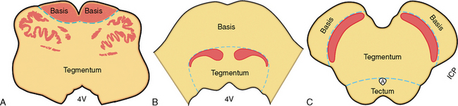
FIGURE 15-5 Ventrodorsal divisions of the brainstem. A, Medulla. B, Pons. C, Midbrain. From ventral to dorsal, the brain stem is divided into three laminae: the basis, the tegmentum, and the tectum. The demarcation between the basis and the tegmentum is the dorsal border of the pyramids (red) in the medulla, the ventral surface of the medial lemnisci (red) in the pons, and the ventral surface of the substantia nigra (red) in the midbrain. The line of demarcation between the tegmentum and the tectum is the ventral aspect of the aqueduct of Sylvius in the midbrain. The medulla and pons have no tectum. 4V, fourth ventricle. In A, the inferior and dorsal accessory olivary nuclei are drawn in to give the tegmentum perspective.
The tegmentum is the middle lamina of the brain stem and also runs its full length. The tegmentum is situated between the basis ventrally and the aqueduct and fourth ventricle dorsally. The tegmentum of the midbrain is defined as the portion of the midbrain between the crura cerebrorum ventrally and the aqueduct dorsally. The tegmentum of the pons is defined as the portion of the pons situated between the medial lemnisci ventrally and the floor of the fourth ventricle. The tegmentum of the medulla is taken to be all of the medulla situated dorsal to the pyramids. The tegmental layer is further subdivided into ventral and dorsal sublaminae. The ventral sublamina contains the supplementary motor nuclei, including the red nuclei and substantia nigra of the midbrain and the inferior olivary nuclei of the medulla. The dorsal sublamina contains multiple cranial nerve nuclei, with the special visceral efferent nuclei situated more ventrally and the general somatic motor and sensory nuclei situated more dorsally (Fig. 15-6). The ventral and lateral portions of the tegmentum also contain the sensory tracts (lemnisci) that ascend to the thalami. The remainder of the tegmentum is occupied by the reticular formation and chemically defined cell groups that produce acetylcholine, norepinephrine, serotonin, and dopamine.
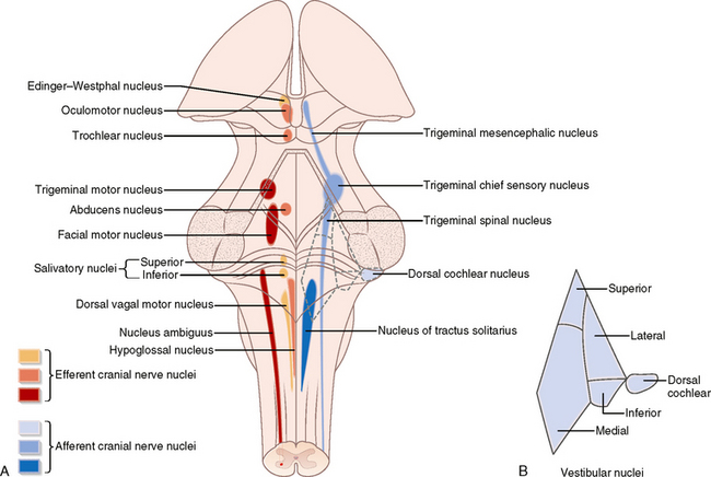
FIGURE 15-6 Cranial nerve nuclei of the brain stem. Projection of the nuclei onto a dorsal schematic of the brain stem.
(From Standring S [ed]. Gray’s Anatomy: The Anatomical Basis of Clinical Practice, 40th ed. Philadelphia, Elsevier, 2008.)
Surface Anatomy
Medulla
Ventral Surface
From medial to lateral on each side, the ventral surface of the medulla displays the anterior median medullary sulcus in the midline, the paired pyramids that flank that sulcus, the paired anterolateral (preolivary) medullary sulci from which the hypoglossal (CN XII) nerve fascicles emerge, the ventral surfaces of the two olives, the paired retro-olivary sulci (lateral fossae of the medulla) from which the fascicles of the glossopharyngeal, vagal, and accessory (CN IX, X, and medullary portion of the XI) nerves emerge, and the ventral surfaces of the two inferior cerebellar peduncles (see Figs. 15-1, 15-2A, and 15-3A).
Lateral Surfaces
From ventral to dorsal on each side, the lateral surface of the medulla displays the lateral aspect of the pyramid, the fascicles of the hypoglossal nerves emerging at the preolivary sulcus, the olive, the retro-olivary sulcus (lateral fossa of the medulla), the supraolivary fossette immediately rostral to the olive and inferior to the middle cerebellar peduncle, the fascicles of CN IX, X, and XI emerging at the retro-olivary sulcus, and the inferior cerebellar peduncle. The lateral funiculus of the spinal cord ascends to join the medulla just behind the olive (see Figs. 15-1, 15-2B, and 15-3C).
Dorsal Surface
The dorsal surface of the medulla is divided into a caudal “closed” portion in which the central canal is covered over as it is in the spinal cord, and a rostral “open” portion in which the central canal opens out into the floor of the fourth ventricle. In the caudal closed portion of the medulla, from medial to lateral on each side, the dorsal surface displays the posterior median medullary sulcus, the paired fasciculi graciles that terminate in the paired gracile tubercles (clavae), the paired posterior intermediate medullary sulci, the fasciculi cuneati that terminate superiorly in the paired cuneate tubercles, the paired posterolateral medullary sulci, and the dorsal surfaces of the two inferior cerebellar peduncles. In the open rostral portion of the medulla, from medial to lateral, the dorsal surface displays the median sulcus, the paired hypoglossal trigones, the paired sulci limitantes, the paired dorsal vagal trigones, the medullary vestibular areas containing portions of the medial and inferior vestibular nuclei,4 and the dorsal aspects of the inferior cerebellar peduncles. The area postrema lies just inferior to the vagal trigone on each side. At the caudal point of the fourth ventricle, the two inferior cerebellar peduncles converge together, creating a shape descriptively termed calamus scriptorius for its resemblance to the sharp cut end of an antique goose quill pen. At this point, the obex of the fourth ventricle leads into the cephalic end of the central canal of the closed portion of the medulla (see Figs. 15-1 and 15-3B).
Pons
Ventrolateral Surface
The curved anterolateral surface of the pons has no distinct ventral or lateral border, so these surfaces are considered together. From medial to lateral on each side, the ventrolateral surface of the pons displays the shallow median pontine sulcus, the horizontally ridged belly of the pons leading directly into the middle cerebellar peduncles, the fascicles of the abducens (CN VI) nerve that exit at the pontomedullary sulcus 1 to 2 cm lateral to the median sulcus, and the fascicles of the facial (CN VII) and vestibulocochlear (CN VIII) nerves that arise at the lateral aspect of the pontomedullary sulcus in the supraolivary fossette (see Figs. 15-1 to 15-3).
Dorsal Surface
The dorsal surface of the pons forms the rostral half of the floor of the fourth ventricle. From medial to lateral on each side, the dorsal surface of the pons (floor of fourth ventricle) displays the median sulcus; the medial eminence; the facial colliculus formed by the passage of the fibers of the facial (CN VII) nerve around and over the nucleus of the abducens (CN VI) nerve; the sulcus limitans; the pontine vestibular area that contains portions of the medial, lateral, and superior vestibular nuclei; the acoustic tubercle that marks the site of the dorsal cochlear nucleus; the trigeminal trigone that marks the site of the principal sensory and motor nuclei of the trigeminal nerve; the dorsal surface of the superior cerebellar peduncle; the parabrachial recess situated between the superior and middle cerebellar peduncles; and the middle cerebellar peduncle most laterally (see Figs. 15-1 and 15-3B).
Mesencephalon (Midbrain)
Ventral Surface
From medial to lateral on each side, the ventral surface of the midbrain displays the median interpeduncular fossa, the fascicles of the oculomotor (CN III) nerve that emerge into the interpeduncular fossa from the medial aspect of each crus cerebri, and the flanking crura cerebrorum. Diencephalic structures seen between the two crura include the paired mammillary bodies, the tuber cinereum, and the pituitary stalk. These hang down into the interpeduncular fossa, but, anatomically, lie outside the confines of the midbrain (see Fig. 15-1).
Lateral Surface
From ventral to dorsal on each side, the lateral surface of the midbrain displays the lateral surface of the crus cerebri, the lateral mesencephalic sulcus, the lateral aspect of the inferior colliculus, the brachium of the inferior colliculus that leads to the medial geniculate body, the superior colliculus, the brachium of the superior colliculus that carries fibers from the optic tract directly to the superior colliculus, bypassing the lateral geniculate nucleus, the exit of the trochlear (CN IV) nerve from the superior medullary velum just caudal to the inferior colliculus, the frenulum veli, and the superior cerebellar peduncle. The pineal gland of the diencephalon overhangs the superior colliculi (see Fig. 15-1).
Dorsal Surface
The dorsal surface of the mesencephalon displays the vertical and horizontal portions of the cruciate intercollicular sulcus, the horizontal infracollicular sulcus caudal to the paired inferior colliculi, the median frenulum veli, the paired superior colliculi, the brachia of the superior colliculi that convey fibers of the optic tract around the lateral geniculate bodies to reach the superior colliculi, the paired inferior colliculi, the brachia of the inferior colliculi that extend to the medial geniculate bodies, the exits of the trochlear (CN IV) nerves just inferior to each inferior colliculus and lateral to the frenulum veli, and the paired lateral mesencephalic sulci (see Figs. 15-1 and 15-4).
Midsagittal Section through the Brain Stem
The midsagittal section through the brain stem displays the central anatomy of the brain stem and the relationship of the brain stem to the cerebellum dorsally and the diencephalon rostrally (see Figs. 15-1 and 15-7). The ventral surface of the brain stem displays the gentle, ventrally directed convexity of the medulla, the inscription point or “notch” at the pontomedullary sulcus, the prominent rounded “belly” of the pons, and the characteristic “7” shape of the interpeduncular fossa of the midbrain. The mammillary bodies of the hypothalamus hang downward into the uppermost portion of the interpeduncular fossa. Farther dorsally, the smooth dorsal convexity of the aqueduct leads downward, along the gentle dorsal concavity of the floor of the fourth ventricle, to the obex, and, via the obex, into the central canal of the closed portion of the medulla. Most dorsally, the superior and inferior colliculi form the roof (tectum) of the midbrain. The fasciculus gracilis of the medulla terminates superiorly at the nucleus gracilis (clava) just behind the obex.
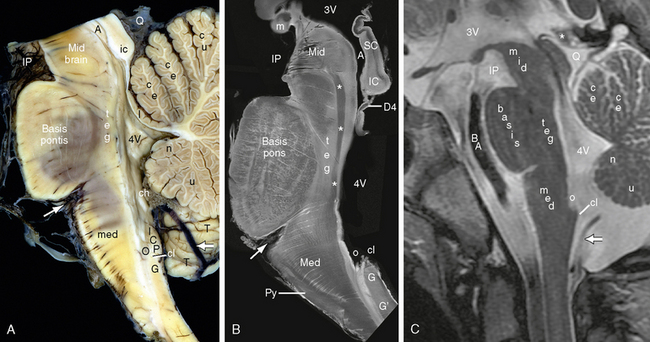
FIGURE 15-7 Midsagittal sections of the brain stem. A, Gross anatomic specimen (see also Fig. 16-4). B, Intermediate-weighted postmortem MR image of the brain stem at 9.4 T (composite image). C, Clinical image at 1.5 T. Note the oblique, curved course of the multiple small vessels that penetrate the ventral aspect of the brain stem. BA, basilar artery traversing the prepontine cistern. Midbrain. The midbrain (mid) shows the characteristic shape of the interpeduncular fossa (IP) ventrally, the aqueduct (A) more dorsally, the superior colliculus (sc) and inferior colliculus (ic) of the quadrigeminal plate (tectum) at the dorsal surface of the brain stem, the quadrigeminal plate cistern (Q) behind the colliculi, and the pineal gland (asterisk in C) that projects into the quadrigeminal plate cistern. In B, the decussation (D4) of the trochlear nerve fascicles appears as a well-defined low signal fiber bundle in the superior medullary velum of the fourth ventricle. The mammillary bodies (m) of the hypothalamus overhang the interpeduncular fossa, contributing to its shape. 3V, third ventricle. Pons. The pons shows the prominent ovoid “belly” formed by the basis pontis, the tegmentum (teg) of the pons situated between the basis pontis and the floor of the upper portion of the fourth ventricle (4V), and the well-defined upper portion of the medial longitudinal fasciculus (white asterisks in B) that extends between the abducens (CN VI) and oculomotor (CN III) nerve nuclei. Medulla. The curved pontomedullary sulcus (oblique white arrow in A and B) delimits the pons from the medulla. The prominent ventral bulge of the basis pontis (“belly” of the pons) is formed by the transversely oriented pontocerebellar fibers. The gently convex curvature of the basis medullae is formed by the pyramids (Py). The dorsal surface of the upper medulla forms the lower half of the floor of the fourth ventricle as far inferiorly as the obex (o). The dorsal surface of the lower closed medulla lies inferior to the fourth ventricle. The fasciculus gracilis (G′) ascends along the dorsal surface of the lower medulla to terminate at the nucleus gracilis (G). The upper pole of the nucleus gracilis forms a dorsal eminence designated the clava (cl). The inferior cerebellar peduncle (ICP) forms the lateral wall of the lower fourth ventricle as it passes superiorly and laterally into the cerebellum. Cerebellum. Behind the brain stem lie the choroid plexus (ch) of the fourth ventricle; the lingula (l) of the vermis attached to the superior medullary velum; the central lobule (ce) and culmen (cu) of the superior vermis; the nodulus (n) and uvula (u) of the inferior vermis; the tonsil (T) and the posterior inferior cerebellar artery (PICA) (thicker horizontal white arrow in A and C). 4V, fourth ventricle.
(B Modified from Naidich TP, Duvernoy HM, Delman BN, et al. Duvernoy’s Atlas of the Human Brain Stem and Cerebellum: High-Field MRI, Surface Anatomy, Internal Structure, Vascularization and 3D Sectional Anatomy. New York, Springer, 2008.)
In parasagittal sections, the medulla displays the pyramid and the start of the pyramidal decussation (see Figs. 15-1 and 15-7). The pons displays the pontine nuclei, the medial lemnisci, the median longitudinal fasciculus, and the penetrating paramedian perforating vessels. The midbrain displays the decussation of the superior cerebellar peduncles and the upper portion of the medial longitudinal fasciculus.
Selected Gross Axial Sections
Gross axial sections display the changing contours of the brain stem, the changing proportions of the basis, tegmentum, and tectum in each portion, and the gross features of the major ascending and descending fiber tracts, gray nuclei, and cranial nerves. These gross anatomic features are illustrated and described in Figures 15-8 to 15-10.
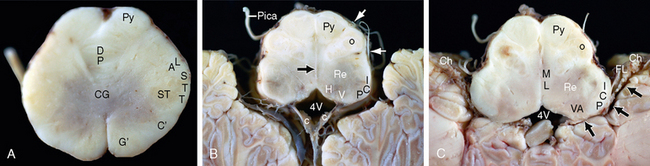
FIGURE 15-8 A to C, Gross axial sections of the medulla displayed from caudal to cranial. Each section is oriented with ventral toward the top of the image. A, Closed far caudal medulla. This section displays the midline ventral sulcus between the two pyramids (Py), the decussation of the pyramids (DP), and the central gray matter (CG). Dorsally and laterally one sees the fasciculi gracilis (G′) and cuneatus (C′), the superficial position of the spinal trigeminal tract (STT), and the spinal trigeminal nucleus (ST) immediately medial to the spinal trigeminal tract. Just ventral to the spinal trigeminal tract, a grouping of ascending and descending fibers forms a composite structure designated the anterolateral fasciculus (AL), which includes the ascending ventral and dorsal spinocerebellar tracts, the ascending spinothalamic tract, and the descending rubrospinal tract. B, Open mid medulla. This section displays the median sulcus and paired pyramids ventrally, the inferior olivary nucleus (O) with ventral preolivary and dorsal retro-olivary sulci (white arrows) laterally, and a thin caudal portion of the inferior cerebellar peduncle (ICP) dorsolaterally. At this level, the floor of the fourth ventricle (4V) shows a narrow transverse dimension with hypoglossal and dorsal vagal eminences that indicate the sites of the underlying hypoglossal (H) and dorsal vagal (V) nuclei. The roof of the inferior fourth ventricle (inferior medullary velum) partially tents over the floor, enclosing the paired choroid plexi (c, c). The median raphe (black arrow), reticular substance (Re), and a branch of the posterior inferior cerebellar artery (PICA) are also indicated. C, Open high medulla. The transverse dimension of the fourth ventricle (4V) is now wider and leads directly into the lateral recesses (three black arrows) of the fourth ventricle on each side. The choroid plexi (Ch) extend the full length of the lateral recesses to emerge from the foramina of Luschka (FL) into the cerebellopontine angle cisterns. The medullary vestibular areas (VA) form the floor of the fourth ventricle at the widest portion of the medullary triangle. In the medulla, the medial lemnisci take the form of paramedian columns of white matter just to each side of the median raphe. Other labels as in A and B.
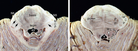
FIGURE 15-9 Pons. A and B, Gross axial sections of the pons displayed from caudal to cranial. Each section is oriented with ventral toward the top of the image. The pons displays an ovoid contour with a gentle ventral median sulcus, a broad lateral extension, and rounded lateral edges. The medial lemniscus (ML) and lateral lemniscus (LL) define the two ends of a lemniscal arc. The ventral surface of this arc marks the interface between the basis pontis (basis) ventrally and the pontine tegmentum (tegm) dorsally. The pontocerebellar fibers (pc) course transversely between the pontine nuclei (p), cross the midline, and gather into the contralateral middle cerebellar peduncles (MCP). The fibers (black arrows) of the trigeminal nerves (N5) penetrate the brain surface in the upper lateral pons and cross through the middle cerebellar peduncles to reach the secondary (relay) trigeminal nuclei in the dorsolateral pons. The superior cerebellar peduncles (SCP) form the lateral wall of the upper fourth ventricle (in A) and then migrate medially into the tegmentum (in B) as they converge to their decussation in the midbrain. On each side, the locus ceruleus (white arrow in B) forms a vertical bluish black column of melanin-containing cells. In cut cross section, these columns appear as small pigmented circles in the dorsal tegmentum.
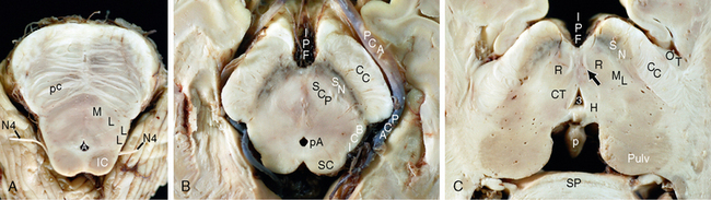
FIGURE 15-10 Midbrain into thalamus. A to C, Gross axial sections from the midbrain into the thalamus displayed from caudal to cranial. Each section is oriented with ventral toward the top of the image. A, Caudal midbrain—upper pons. The caudal midbrain shows a narrow transverse extension and gently rounded contours. In the tegmentum, the lateral lemnisci (LL) contribute to the capsule of white matter surrounding the inferior colliculi (IC). The trochlear nerves (N4) emerge from the dorsal surface (not seen) and then normally course ventrally around the low midbrain. In this image, the cut ends of the two trochlear (N4) nerves pass artifactually far laterally. The ventral aspect of the lemniscal arc (ML-LL) divides the basis ventrally from the rest of the tegmentum dorsally. B, Mid midbrain. The mid midbrain shows a wider transverse dimension, more angular contours, and a deep interpeduncular fossa (IPF) containing multiple posterior thalamoperforating vessels. The anterior border of the substantia nigra (SN) separates the crura cerebrorum (CC) ventrally from the tegmentum of the midbrain dorsally. The aqueduct (unlabeled) separates the tegmentum ventrally from the tectum dorsally. The uppermost portions of the superior cerebellar peduncles (SCP) are still visible within the tegmentum and will decussate one level more superiorly. The brachium of the inferior colliculus (BCI) passes ventral to the superior colliculus (SC) en route to the medial geniculate body. The posterior cerebral artery (PCA) encircles the midbrain. pA, periaqueductal gray matter. C, Upper midbrain. The optic tract (OT) encircles the crus cerebri (CC) on each side. The tegmentum displays the substantia nigra (SN), the paired red nuclei (R), the central tegmental tracts (CT) at the dorsomedial aspects of the red nuclei, and the medial lemnisci (ML) at the dorsolateral aspects of the red nuclei. The paired habenulointerpeduncular tracts (fasciculi retroflexi) (black arrow) pass ventrally from the habenular trigones (H) at the posterior third ventricle through the medial aspects of the red nuclei to reach the interpeduncular nuclei. Other structures labeled include the third ventricle (3), pineal gland (P), pulvinar (pulv), and splenium (Sp).
Selected Axial Histologic Sections
Lower Medulla
Axial sections through the caudal (closed) medulla (Fig. 15-11A and B) display the central canal and the central gray matter surrounding the canal. From ventral to dorsal on each side, the central gray matter contains the nuclei of the hypoglossal nerve (CN XII), the dorsal motor nucleus for the vagus nerve (CN X), and the nucleus of the solitary tract. From medial to lateral, the dorsal quadrant displays the fasciculus and nucleus gracilis, the fasciculus and nucleus cuneatus, and the spinal trigeminal tract and nucleus. In this plane, the spinal trigeminal tract lies at the lateral surface of the medulla. Continuing around the curvature, from lateral to medial, the anterior quadrant of the lower medulla displays the site of the anterolateral fasciculus and the pyramidal tract. At the caudal medullary level, the anterolateral fasciculus contains the dorsal and ventral spinocerebellar tracts at the surface and the spinothalamic tract just deep to those. A rubrospinal tract is seen within the anterolateral fasciculus in other species but is rudimentary in humans.
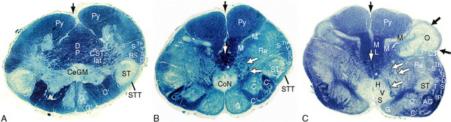
FIGURE 15-11 A to C, Axial histologic sections of the lower medulla. Figures 15-11 to 15-13 are histologic sections displayed in ascending order from caudal to rostral. Each section is oriented with ventral toward the top of the image. Klüver-Barrera stain for myelin (blue) and gray matter nuclei purplish). The inferior medulla lies caudal to the fourth ventricle. Far caudally (A), the medullary gray matter resembles the spinal cord with the central gray matter (CeGM) surrounding the closed central canal, and ventral gray matter (white V) reminiscent of ventral horns far laterally. Farther rostrally (B), the central gray matter contains the commissural nucleus (CoN) through which the caudal ends of the paired nuclei of the solitary tracts unite in the midline. Near the top of the closed medulla (C), the central gray matter moves dorsally and displays a vertical alignment of nuclei, with the darker-stained more myelinated hypoglossal nucleus (H) situated ventrally, the lighter less myelinated dorsal nucleus of the vagus (V) situated centrally, and the light nucleus of the solitary tract (S) situated most dorsally. Dorsally, the dorsal sensory fasciculi gracilis (G′) and cuneatus (C′) ascend to their relay nuclei gracilis (G) and cuneatus (C). From these nuclei, secondary relay fibers designated the internal arcuate fibers (white arrows) curve ventrally around the central gray nuclei, cross the midline ventrally as the lemniscal (great sensory) decussation (thin vertical white arrows), and emerge on the contralateral side to form the paired medial lemnisci (ML). In the lower and mid medulla, the medial lemnisci form paired paramedian columns of white matter. The accessory cuneate nucleus (AC) first appears as small islands of gray matter within the upper fasciculus cuneatus (also seen, unlabeled, between the two C′ in B). In more rostral sections (C and above), the accessory cuneate nucleus becomes a substantial nucleus lying rostrolateral to the cuneate nucleus. The medial longitudinal fasciculi (white asterisk) form a vertically oriented fiber system that courses in the general area ventral to the central gray matter and dorsal to the medial lemnisci (ML). Ventrally, the corticospinal tracts descend within the paired paramedian pyramids (Py) to each side of the anterior median medullary sulcus (thin vertical black arrow). They decussate (DP) in the lower medulla and then course posteroinferolaterally (CST lat) toward the posterior aspect of the lateral columns of the cord (caudal to the sections shown). Laterally, the descending fibers of the spinal trigeminal tract (STT) present at the surface of the medulla inferiorly (A and B) but become buried deep to the inferior cerebellar peduncle (IP) more rostrally. The large spinal trigeminal nucleus (ST) runs vertically directly medial to the spinal trigeminal tract. The dorsal spinocerebellar tract (DS) and ventral spinocerebellar tract (VS) lie adjacent to each other as they ascend into the low medulla (A). However, the dorsal spinocerebellar tract then separates from the ventral spinocerebellar tract and curves dorsally as it ascends to enter the inferior cerebellar peduncle (IP) at more rostral levels (B and C). Just anterolateral to the spinal trigeminal system, multiple fiber systems course vertically in a combined anterolateral fasciculus. These anterolateral fibers include the ascending ventral spinocerebellar tract (VS) and the ascending spinothalamic tract (STh). The inferior olivary nucleus (O) and its capsule (the amiculum, two black arrows) form the gross bulge designated the olive. Between the inferior olivary nucleus and the medial lemniscus, the medial accessory olivary nucleus (M) forms a vertical column of gray with a ventral horizontal wing oriented at right angles to the vertical dorsal wing. Also indicated are the accessory cuneate nucleus (AC) and the reticular formation of the medulla (one portion of which is labeled Re).
Mid Medulla
Axial sections through the upper end of the closed portion of the medulla (see Fig. 15-11C) display anatomy similar to that just described. Centrally, however, the secondary relay (internal arcuate) fibers arise from the dorsal nuclei gracilis and cuneatus and pass ventrally around the central gray matter to decussate in the ventral midline. The decussated fibers assemble into compact fiber bundles just to each side of the midline and are renamed the medial lemnisci. Dorsally, the accessory cuneate nucleus now appears lateral to the cuneate nucleus. Laterally, the dorsal spinocerebellar tract has moved dorsally in preparation to enter the inferior cerebellar peduncle more rostrally. As a result, the spinal trigeminal tract and nucleus now lie deep to the dorsal spinocerebellar tract and the anterolateral fasciculus now contains the ventral spinocerebellar tract and the spinothalamic tract but not the dorsal spinocerebellar tract. The medial accessory olivary nucleus may be seen within the ventral quadrant.
Upper Medulla
Axial sections through the rostral (open) medulla (Fig. 15-12) display the lower half of the fourth ventricle. The central gray matter that surrounded the central canal more caudally now opens dorsally and folds outward, so the previously ventral nuclei now lie medially and the previously dorsal nuclei now lie laterally. From medial to lateral, therefore, the medullary portion of the floor of the fourth ventricle displays the hypoglossal nucleus at the hypoglossal trigone, the dorsal nucleus of the vagus at the vagal trigone, and the nucleus of the solitary tract lateral to the vagal nucleus and extending deeply to surround the solitary tract. The medullary vestibular area containing the medial and inferior vestibular nuclei lies at the lateral end of the floor of the fourth ventricle. The ventral surface of the rostral medulla displays the paired paramedian pyramids carrying the corticospinal tracts, the preolivary sulci at which the hypoglossal (CN XII) nerve roots emerge, the olives formed by the underlying inferior olivary nuclei, and the retro-olivary sulci at which the fibers of the glossopharyngeal, vagus, and accessory nerves (CN IX, X, and XI) transit into and out from the stem. The dorsal quadrant displays the nucleus ambiguus and the inferior cerebellar peduncle (restiform body) that now contains the ipsilateral dorsal spinocerebellar fibers and the olivocerebellar fibers that arise from the contralateral inferior olivary nucleus. The anterolateral fasciculus lies along the surface of the upper medulla just ventral to the inferior cerebellar peduncle. The ventral quadrant displays the C-shaped, markedly folded inferior olivary nucleus and the medial accessory and dorsal accessory olivary nuclei. Just to each side of the midline, from dorsal to ventral, rostral medullary sections display the paired medial longitudinal fasciculi and the paired medial lemnisci.
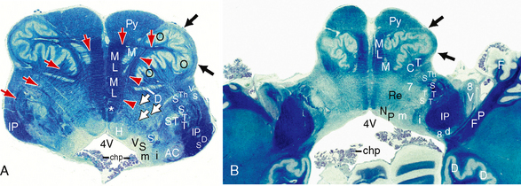
FIGURE 15-12 A and B, Axial histologic sections of the upper medulla. Figures 15-11 to 15-13 are histologic sections displayed in ascending order from caudal to rostral. Each section is oriented with ventral toward the top of the image. Klüver-Barrera stain for myelin (blue) and gray matter nuclei (purplish). The rostral medulla displays the lower half of the open fourth ventricle (inferior medullary triangle of the rhombencephalic ventricle) (4V). A, The central canal opens dorsally, so the gray matter nuclei that were situated dorsally in the low medulla come to lie laterally in the upper medulla. Dorsally, along the floor of the open fourth ventricle, the hypoglossal nuclei (H) lie medially and bulge into the floor of the ventricle at the hypoglossal trigones. The more lightly myelinated dorsal vagal nuclei (V) lie lateral to these and bulge into the floor of the fourth ventricle at the vagal trigones. The nuclei (black S) of the solitary tract lie lateral to the vagal nuclei and extend ventrolaterally to surround the myelinated solitary tracts (s). The vestibular nuclei form the vestibular areas at the lateral aspects of the fourth ventricle. In this section, these include the medial (black m) and the inferior (i) vestibular nuclei. The spinal trigeminal nucleus (ST) and tract (STT) occupy nearly the same location but at this level are buried deeply by the expansion of the inferior cerebellar peduncle (IP). Internal arcuate fibers (white arrows) continue to course from the nuclei gracilis and cuneatus across the midline into the contralateral medial lemnisci. Ventrally, the paired pyramids (Py) lie to each side of the anterior median medullary sulcus. Laterally, the dorsal spinocerebellar tract has moved farther into the inferior cerebellar peduncle (IP). The ascending ventral spinocerebellar tract (VS) and the ascending spinothalamic tract (STh) maintain their positions within the anterolateral fasciculus. The inferior olivary nucleus (O) and its capsule (amiculum, black arrows) bulge laterally, creating the preolivary sulcus and retro-olivary sulcus. Large numbers of olivocerebellar fibers (red arrows) emerge from the hilum of one olive, pass medially to cross the midline, and then turn dorsally and laterally to enter the contralateral inferior cerebellar peduncle (IP). In their course, they cross directly through all intervening structures, including the medial accessory olive (white

Stay updated, free articles. Join our Telegram channel

Full access? Get Clinical Tree


