CHAPTER 17 Cranial Nerves
The twelve cranial nerves consist of the olfactory (I), optic (II), oculomotor (III), trochlear (IV), trigeminal (V), abducens (VI), facial (VII), vestibulocochlear (VIII), glossopharyngeal (IX), vagus (X), accessory (XI), and hypoglossal (XII) nerves. Each nerve has an intra-axial, cisternal, dural, osseous, and extracranial segment. This chapter addresses the anatomy and function of the cranial nerves and the different imaging techniques preferred for depicting each segment of the cranial nerves.
GROSS ANATOMY
From cranial to caudal, cranial nerves II to XII can be grouped by their origin from the brain stem:
• CN II originates from the diencephalon.
• CN III and IV originate from the mesencephalon.
• CN V through VIII originate from the pons.
CN I: Olfactory Nerve, Bulb, and Tract
The olfactory nerve consists of a collection of sensory nerve fibers (the olfactory filiae), which represent the axons of the olfactory receptor neurons, located in the olfactory mucosa of the nasal cavity. The olfactory filiae enter the anterior cranial fossa via the cribriform plate of the ethmoid bone (Fig. 17-1) and terminate in the olfactory bulbs, which lie in bony grooves formed by the cribriform plate (see Figs. 17-1 and 17-2). The olfactory bulbs constitute the primary olfactory cortices. In the olfactory bulbs, the primary axons form synapses with the secondary neurons of the olfactory system. The axons of the secondary neurons leave the olfactory bulb in the olfactory tract, which runs inferior to the olfactory sulcus (see Fig. 17-2) to reach the posterior aspect of the orbital surface of the forebrain. At the rostral border of the anterior perforated substance the olfactory tract divides into three olfactory striae: (1) the medial olfactory stria, which curves medially and upward to the septal region, (2) the lateral olfactory stria, which first curves far laterally along the sylvian fissure and then recurves medially to reach the medial surface of the temporal lobe, and (3) the vestigial intermediate olfactory stria, which continues onto the anterior perforated substance and ends at the olfactory tubercle. The striae and tubercle constitute the olfactory trigone. Areas that receive direct projections from the olfactory tract consist of the anterior olfactory nucleus, the olfactory tubercle, the piriform cortex, the anterior cortical nucleus of the amygdala, the periamygdaloid cortex, and a small anteromedial portion of the entorhinal cortex. These constitute the secondary olfactory cortices.
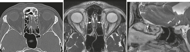
FIGURE 17-1 Axial (A, B) and sagittal (C) scans through the olfactory groove. A, CT. B and C, MR FIESTA sequence. 1, ethmoid plate; 2, inferior orbital fissure; 3, olfactory bulb.
CN II: Optic Nerve, Chiasm, and Tract
The optic nerve emerges from the posterior pole of the ocular globe, extends posteriorly through the orbit, and leaves the orbit via the optic canal to reach the anterior cranial fossa (Fig. 17-3). Intracranially, both optic nerves join to form the optic chiasm within the suprasellar cistern. At the chiasm the nasal fibers of each optic nerve cross the midline (decussate) to join with the uncrossed temporal fibers of the opposite optic nerve, forming the optic tracts (see Fig. 17-3). From the optic chiasm, the paired optic tracts curve posteriorly around the cerebral peduncles to terminate in the lateral geniculate bodies (see Fig. 17-3). From the lateral geniculate bodies the optic radiations curve toward the primary visual cortices (Brodmann areas 17) at the medial aspect of the occipital lobes, along the calcarine and anterior calcarine fissures.
CN III: Oculomotor Nerve
The nuclear complex of the third nerve is located in the midbrain at the level of the superior colliculi immediately dorsal to the medial longitudinal fasciculus (MLF) and just ventral to the periaqueductal gray matter (Fig. 17-4). The parasympathetic motor fibers originate in the Edinger-Westphal nucleus situated at the dorsal-rostral aspect of the oculomotor nuclear complex. The fascicles of CN III curve ventrally through the midbrain at the level of the red nucleus and exit the brain stem along the ventromedial aspect of the cerebral peduncle within the interpeduncular fossa (see Fig. 17-4). The cisternal segment of CN III courses through the interpeduncular and the perimesencephalic cisterns, passing between the P1 segment of the posterior cerebral artery (PCA) above and the superior cerebellar artery (SCA) below (see Fig. 17-4). CN III then enters the lateral dural wall of the cavernous sinus and courses superiorly and laterally within it (Fig. 17-5) to enter the orbital cavity via the superior orbital fissure within the annulus of Zinn (the common tendinous ring of the extraocular muscles).
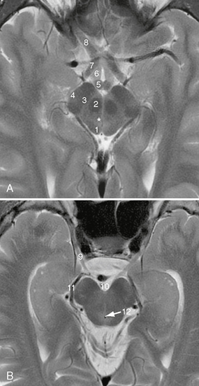
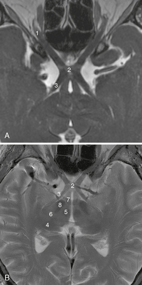
FIGURE 17-4 A and B, Axial T2W MR images at the level of the midbrain. 1, superior colliculus; 2, red nucleus; 3, substantia nigra; 4, cerebral peduncle; 5, mammillary body; 6, fornix; 7, optic chiasm; 8, optic nerve; 9, oculomotor nerve; 10, interpeduncular fossa; 11, posterior cerebral artery; 12, cerebral aqueduct. Asterisk is on the oculomotor nucleus.
CN IV: Trochlear Nerve
The nucleus of CN IV is located in the midbrain at the level of the inferior colliculus, caudal to the nuclear complex of CN III, immediately dorsal to the medial longitudinal fasciculus (MLF), and just ventral to the periaqueductal gray matter (Fig. 17-6). The nerve fascicles pass posteriorly and caudally within the periaqueductal gray matter and then decussate within the superior medullary velum before emerging at the dorsal surface of the midbrain just inferior to the contralateral inferior colliculus (see Fig. 17-6). CN IV is the sole cranial nerve to exit the brain stem dorsally and on the side opposite its nucleus. CN IV then courses ventrally and laterally through the quadrigeminal and ambient cisterns to reach the free margin of the tentorium cerebelli, where it typically runs just inferior and lateral to the free margin of the tentorium cerebelli (see Fig. 17-6).1 The trochlear nerve enters the cavernous sinus just inferior to the petroclinoid ligament and passes anteriorly in the lateral dural wall of the cavernous sinus, inferior to the third nerve and superior to the ophthalmic division of the fifth cranial nerve (see Fig. 17-5). After passing the cavernous sinus, it enters the orbital apex via the superior orbital fissure external to the annulus of Zinn to innervate the contralateral superior oblique muscle.
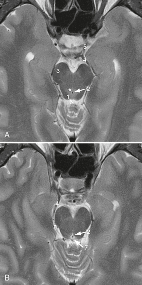
FIGURE 17-6 A and B, Axial T2W MR images at the level of the inferior colliculi. Asterisk, trochlear nucleus; 1, inferior colliculus; 2, cerebral aqueduct; 3, cerebral peduncle; 4, posterior cerebral artery; 5, posterior communicating artery; 6, quadrigeminal cistern; 7, superior medullary velum; 8, ambient cistern.
CN V: Trigeminal Nerve
CN V consists of preganglionic roots, a large sensory root and one to three smaller motor roots,2 as well as three postganglionic branches: the ophthalmic nerve (CN V1), the maxillary nerve (CN V2), and the mandibular nerve (CN V3).
The trigeminal ganglion (also called the semilunar or gasserian ganglion) is situated along the anterior inferior lateral wall of Meckel’s cave. Meckel’s cave itself is formed by the meningeal layer of the dura and is located within the middle cranial fossa posteroinferolateral to the cavernous sinus. It is largely filled with cerebrospinal fluid and houses the trigeminal ganglion at its inferior aspect (Fig. 17-7).
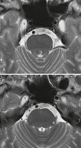
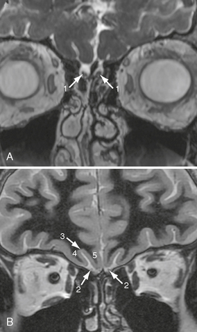
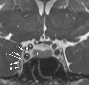
FIGURE 17-7 A and B, Axial T2W MR images at the level of the superior cerebellar peduncles. 1, Meckel’s cave; 2, trigeminal nerve roots; 3, medial longitudinal fasciculus; 4, point of exit/entry of trigeminal nerve roots; 5, fourth ventricle; 6, middle cerebellar peduncle (brachium pontis); 7, superior cerebellar peduncle (brachium conjunctivum); 8, anterior inferior cerebellar artery (AICA).
The trigeminal sensory nerve root emerges from the concave portion of the trigeminal ganglion as several small sensory rootlets that pass posteriorly to enter the brain stem at the level of the lateral pons (see Fig. 17-7). The motor nerve roots of CN V exit the pons just anterosuperomedial to the entry point of the sensory root.2 All CN V nerve roots traverse the prepontine cistern in close relationship to the superior cerebellar artery (see Fig. 17-7) and the anterior inferior cerebellar artery (AICA). The motor root of CN V passes through Meckel’s cave inferior to the trigeminal ganglion and is adherent to the basal wall of the cave in its distal portion.3
The postganglionic branches of CN V (CN V1-V3) emerge from the convex portion of the trigeminal ganglion. CN V1 and CN V2 run through the lateral wall of the cavernous sinus inferior to CN IV (see Fig. 17-5).4 The ophthalmic nerve (CN V1) then enters the orbit via the superior orbital fissure together with CN III, CN IV, and CN VI and the superior ophthalmic vein. The nasociliary branch of CN V1 passes through the annulus of Zinn, while the lacrimal and frontal branches of CN V1 enter the orbit outside the annulus of Zinn. CN V2 leaves the skull base through the foramen rotundum (Fig. 17-8) and crosses the pterygopalatine fossa. The nerve then courses laterally, enters the orbit via the inferior orbital fissure (see Fig. 17-8), and passes through (or beneath) the orbit within the infraorbital groove/canal to reach the face via the infraorbital foramen.
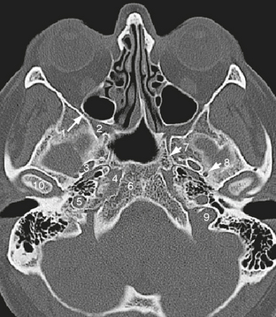
FIGURE 17-8 Axial CT scan at the level of the inferior orbital fissure. 1, inferior orbital fissure; 2, foramen rotundum; 3, foramen ovale; 4, foramen lacerum; 5, carotid canal, vertical portion; 6, basioccipital portion of clivus; 7, vidian canal; 8, foramen spinosum; 9, jugular foramen, pars vascularis; 10, mandibular condyle.
The third division of CN V, the mandibular nerve, consists of the inferior division of the sensory root and the motor root. These nerve roots leave the skull base through the foramen ovale (see Fig. 17-8), unite, and run through the infratemporal fossa. Within the infratemporal fossa, CN V3 gives off some branches and then divides into an anterior and a posterior division, both of which give off multiple branches (e.g., to the muscles of mastication, the otic ganglion, the skin of the temporal region, the auricle, and the buccal mucosa).
CN VI: Abducens Nerve
The nucleus of the abducens nerve lies just beneath the floor of the fourth ventricle in the dorsal pons, close to the medial longitudinal fasciculus (MLF). The fascicles of the seventh nerve course around the abducens nucleus, first medial, then dorsal, then lateral to it, forming a protuberance that bulges into the fourth ventricle as the facial colliculus (Fig. 17-9). The abducens fibers course ventrally, caudally, and slightly laterally to exit the brain stem at the pontomedullary sulcus (or the caudalmost pons). The cisternal segment of CN VI recurves anteriorly, laterally, and rostrally; approaches the upper clivus; and passes through a dural channel within the basilar venous plexus (Dorello’s canal)5 to enter the cavernous sinus (see Fig. 17-5). Within the cavernous sinus, CN VI courses along the lateral aspect of the internal carotid artery and its surrounding sympathetic plexus (see Fig. 17-5). CN VI is the only cranial nerve to actually course through the central venous portion of the cavernous sinus. CN III, CN IV, CN V1, and CN V2 all course in the lateral dural wall of the cavernous sinus, not within the central venous portion. CN VI then enters the orbit via the superior orbital fissure within the annulus of Zinn.
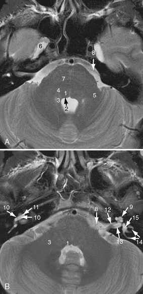
FIGURE 17-9 A and B, Axial T2W MR images at the level of the facial colliculi. 1, abducens nucleus; 2, facial colliculus (genu of facial nerve); 3, superior cerebellar peduncle (brachium conjunctivum); 4, facial nucleus; 5, middle cerebellar peduncle (brachium pontis); 6, Meckel’s cave; 7, pontocerebellar fibers; 8, anterior inferior cerebellar artery (AICA); 9, second turn of cochlea; 10, basal turn of the cochlea, scala vestibuli, 10′, basal turn of the cochlea, scala tympani; 11, osseous spiral lamina; 12, cochlear nerve; 13, inferior vestibular nerve; 14, lateral and posterior semicircular canals; 15, vestibule.
CN VII: Facial Nerve
CN VII consists of two portions: (1) CN VII proper, a pure motor nerve, and (2) the intermediate nerve (nervus intermedius), which contains both sensory and parasympathetic motor fibers (see later). Both portions merge at the geniculate ganglion (see later).
The motor nucleus of CN VII is located in the lower portion of the pons, ventral, lateral and slightly caudal to the nucleus of CN VI (see Fig. 17-9). On each side, the motor fasciculi of CN VII course dorsally and cranially and pass medial to the abducens nucleus, and then dorsal to it, between the floor of the fourth ventricle and the abducens nucleus to form the facial colliculus along the floor of the fourth ventricle (see Fig. 17-9). The fascicles of CN VII then curve ventrally and laterally to exit the ventral brain stem at the lower border of the pons in the lateral portion of the pontomedullary sulcus (medial to CN VIII). CN VII traverses the cerebellopontine angle cistern to enter the petrous portion of the temporal bone via the internal acoustic meatus (see Fig. 17-9 for details on the intrameatal course and see the later section on CN VIII).
CN VII runs through the petrous bone within the facial canal, which extends from the internal acoustic meatus to the stylomastoid foramen (Fig. 17-8).6 Three portions of CN VII can be distinguished within the facial canal:
1. The first segment (labyrinthine portion) begins in the internal acoustic meatus and runs perpendicular to the long axis of the petrous bone, directed anteriorly and laterally.
2. The second segment (tympanic portion) begins at the geniculate ganglion and curves dorsally and slightly caudally, between the lateral semicircular canal (superiorly) and the oval window (inferiorly).
3. The third segment (mastoid portion) runs vertically toward the stylomastoid foramen (see Fig. 17-14).
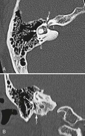
FIGURE 17-14 Axial CT scan (A) and coronal reconstruction (B) through the right temporal bone. 1, Internal acoustic meatus; 2, vestibule; 3, tympanic segment of facial canal; 4, lateral semicircular canal; 5, posterior semicircular canal; 6, mastoid segment of facial canal; 7, stylomastoid foramen.
The geniculate ganglion is located at the distal end of the labyrinthine portion. The cell bodies of the afferent fibers of the intermediate nerve are located within this ganglion. These afferent fibers originate from receptors within the first two thirds of the tongue. They first course with the lingual nerve (CN V) to reach the geniculate ganglion via the chorda tympani (see later). They then run with the intermediate nerve through the internal acoustic meatus and the cerebellopontine angle cistern (Fig. 17-10) to terminate in the upper pole of the nucleus of the solitary tract (designated the gustatory nucleus).
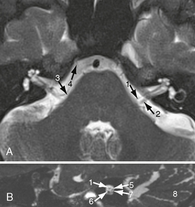
FIGURE 17-10 Axial (A) and sagittal (B) scans though the internal acoustic meatus. A, MR T2W image. B, MR FIESTA sequence. 1, Facial nerve; 2, vestibulocochlear nerve; 3, anterior inferior cerebellar artery (AICA); 4, abducens nerve; 5, superior vestibular nerve; 6, cochlear nerve; 7, inferior vestibular nerve; 8, cerebellum.
CN VIII: Vestibulocochlear Nerve
CN VIII consists of two components: the cochlear (or auditory) nerve and the vestibular nerve.
The cochlear and the vestibular nerves traverse the internal acoustic meatus to reach the cerebellopontine angle cistern. Within the internal acoustic meatus, CN VII, the superior and the inferior vestibular nerve, as well as the cochlear nerve run parallel to each other, with CN VII anterosuperior, the superior vestibular nerve posterosuperior, the cochlear nerve anteroinferior, and the inferior vestibular nerve posteroinferior (see Fig. 17-10).
CN IX: Glossopharyngeal Nerve
The motor fibers of CN IX originate in the nucleus ambiguus. The parasympathetic fibers originate in the inferior salivary nucleus. The sensory fibers of CN IX project to the solitary nucleus and the spinal nucleus of the trigeminal nerve. These nuclei all reside in the medulla oblongata. CN IX emerges from the medulla oblongata as 10 to 20 rootlets along the upper third of the postolivary sulcus.7,8 These rootlets form the cisternal segment of CN IX and traverse the lateral cerebellomedullary cistern (Fig. 17-11) to enter the jugular foramen through the glossopharyngeal meatus (see Fig. 17-11). Within the jugular foramen, CN IX forms a superior and an inferior ganglion. The superior ganglion is located immediately below the external opening of the cochlear aqueduct, and the inferior ganglion is located immediately caudal to the superior one.8
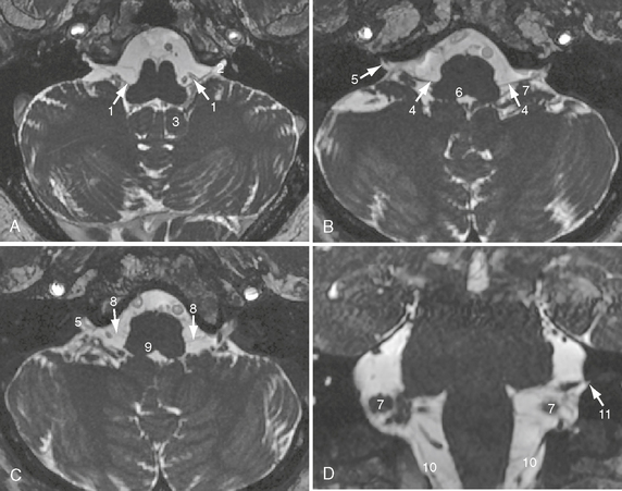
FIGURE 17-11 Axial (A–C) and coronal (D) images at the level of the medulla oblongata. MR FIESTA sequence. 1, Glossopharyngeal nerve; 2, glossopharyngeal meatus; 3, cerebellar tonsil; 4, vagus nerve; 5, vagal meatus; 6, vagal trigone; 7, flocculus; 8, cranial nerve roots of the accessory nerve; 9, hypoglossal trigone; 10, spinal root of the accessory nerve; 11, external opening of the cochlear aqueduct.
At the inferior ganglion, CN IX gives off its tympanic branch (Jacobson’s nerve), which traverses the tympanic canaliculus to enter the tympanic cavity.8
CN X: Vagus Nerve
The motor fibers of CN X emerge from the base of the nucleus ambiguus and from the dorsal nucleus of the vagus (i.e., the upper portion of the spinal nucleus of the vagus). Both nuclei are located in the medulla oblongata, where the dorsal nuclei of CN X form the “vagal trigone” at the caudal aspect of the floor of the fourth ventricle (Fig. 17-11). The CN X rootlets emerge from the brain stem along the upper third of the postolivary sulcus, slightly caudal to CN IX, and typically form two nerve bundles (the upper and lower CN X nerve root bundles).8,9 These traverse the lateral cerebellomedullary cistern and enter the jugular foramen via the vagal meatus (see Fig. 17-11), which is located immediately inferior to the glossopharyngeal meatus. CN X forms a superior ganglion at the level of the intracranial orifice of the jugular foramen. This extends below the extracranial orifice of the jugular foramen. The auricular branch of CN X (Arnold’s nerve) emerges from CN X at this ganglion. CN X traverses the jugular foramen in the immediate vicinity of CN XI and posteromedial to CN IX (Fig. 17-12). Just below the jugular foramen, CN X forms the inferior vagal ganglion and runs in close relationship to CN XII, which courses medially to CN X.7
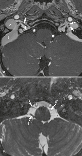
FIGURE 17-12 Axial scans at the level of the hypoglossal canal. A, T1W contrast-enhanced MR image. B, MR FIESTA sequence. 1, Internal carotid artery; 2, jugular bulb within the venous portion of jugular foramen; 3, sigmoid sinus; 4, hypoglossal canal; 5, hypoglossal nerve, canalicular segment; 5′, hypoglossal nerve, cisternal segment; 6, glossopharyngeal nerve, jugular foramen portion; 7, vagus nerve, jugular foramen portion; 8, posterior inferior cerebellar artery (PICA).
CN XI: Spinal Accessory Nerve
CN XI is traditionally considered to have both a cranial root (crCN XI) and a spinal root (spCN XI). However, the existence of the cranial roots of CN XI is controversial. Some authors assign the “traditional” crCN XI rootlets to the vagus nerve, because there is evidence that these rootlets originate in the caudal portion of the nucleus ambiguus and the dorsal nucleus of the vagus nerve. Thus they are functionally vagal rootlets.10,11 Six to 16 of these so-called crCN XI rootlets emerge from the medulla oblongata in the retro-olivary sulcus, just caudal to the vagal nerve bundles (see Fig. 17-11).

Stay updated, free articles. Join our Telegram channel

Full access? Get Clinical Tree


