Breast Biopsy

5 Breast Biopsy


Jennifer A. Harvey

Classification


Percutaneous image-guided breast biopsy is less invasive, less expensive, and more efficient than diagnostic surgical biopsy.


  • Sensitivity of ultrasound-guided breast core needle biopsy (CNB): 97–9%
  • Fine-needle aspiration biopsy (FNAB) is less sensitive, but can be very useful in evaluating additional lesions or abnormal lymph nodes in the setting of a known primary breast cancer.
  • Most breast masses are amenable to ultrasound-guided biopsy.

Initial Training


American College of Radiology (ACR) Practice Guidelines for performance of ultrasound-guided breast biopsy recommends that radiologists who are beginning to perform these procedures meet practice guidelines for performing breast ultrasound and obtain


  • At least 3 hours of continuing professional education hours in breast ultrasound intervention, and perform at least three procedures under direct supervision. These training suggestions can be met in residency or fellowship training programs.
  • In addition, the ACR recommends that radiologists perform at least 12 ultrasound-guided procedures annually to maintain skills.

Advantages of image-guided preoperative diagnosis of breast cancer over diagnostic surgical biopsy:


  • Reduces the number of women undergoing surgical procedures
  • Allows the patient and surgeon to discuss therapeutic options in advance of surgery
  • Once a breast cancer has been diagnosed, the extent of disease can be evaluated by ultrasound or magnetic resonance imaging (MRI). Ultrasound-guided biopsy documenting multifocal or multicentric disease may alter surgical management.

    • Percutaneous biopsy of nonpalpable cancer results in a single therapeutic surgical operation in 81% of women. Receptor status as determined by CNB samples can also guide neoadjuvant chemotherapy if needed.

Other advantages of ultrasound guidance for breast CNB over stereotactic guidance include


  • The lack of ionizing radiation is particularly important for pregnant women with a breast mass.
  • Real-time visualization of the needle during biopsy improves confidence that a small lesion has been accurately sampled.
  • The supine position of the patient for an ultrasoundguided procedure is often more comfortable than the prone or sitting position used for stereotactic procedures.
  • Stereotactic biopsy is also limited by compressed breast thickness of at least 2.5 cm, whereas small breast thickness rarely affects the ability to perform ultrasound-guided breast biopsy.
  • Ultrasound-guided breast biopsies (in experienced hands) are often more efficient than stereotactic biopsy.

Indications


Percutaneous image-guided biopsy has largely replaced diagnostic surgical biopsy in the evaluation of breast lesions in many practices. Most breast lesions that are visible on ultrasound that are suspicious (Breast Imaging Reporting and Data System [BI-RADS] assessment category 4) or malignant (BI-RADS 5) are amenable to ultrasoundguided core needle biopsy. Image-guided biopsy is not typically performed for


  • Lesions with BI-RADS assessment category 3: Probably benign, unless there is undue patient anxiety or the reliability of the patient for return visit is uncertain, for example a homeless woman with limited healthcare resources.
  • In the setting of a palpable finding, diagnostic ultrasound-guided biopsy can be an alternative to excision.

Some lesions will not be amenable to percutaneous biopsy. Of 630 breast cancers diagnosed over a 3-year period at Yale University, 17% were not diagnosed by percutaneous biopsy. The most commonly cited reasons were


  • Lesion was difficult to visualize for image-guided biopsy.
  • Patient preference
  • Lesion was superficial or very small.

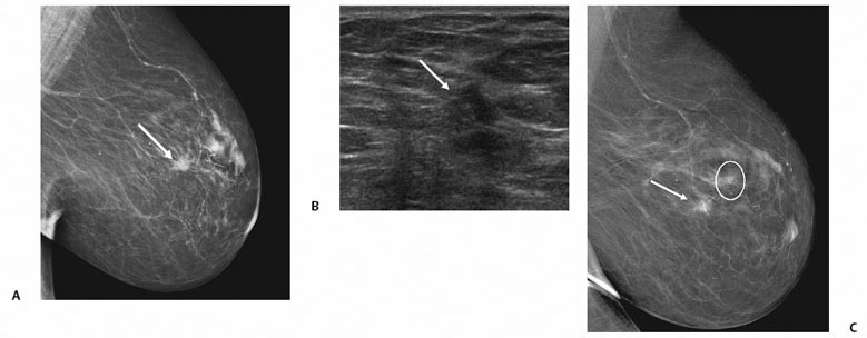
Although most masses identified on mammography can be identified on ultrasound for biopsy, lesions presenting as architectural distortion or calcifications on mammography may be more difficult to identify on ultrasound. If a lesion is much better visualized on mammography than ultrasound, then stereotactic-guidance may be the better choice to improve confidence that the correct area was sampled (Fig. 5.1).


image


Fig. 5.1 A 67-year-old woman with abnormal right mammogram. (A) Right mediolateral (ML) view shows an 8-mm mass with spiculated margins (arrow). (B) Ultrasound shows a corresponding subtle hypoechoic solid mass in the right breast at 10 o’clock (arrow). (C) Postprocedure ML view shows that the clip (circle) is located ~2 cm from the lesion (arrow). Core biopsy was immediately repeated using stereotactic guidance. Histology from ultrasoundguided core needle biopsy (CNB) showed only stromal fibrosis, whereas the stereotactic-guided CNB showed ductal carcinoma in situ. In retrospect, stereotactic-guided CNB would likely have been a better choice initially because the finding was subtle on ultrasound.


Microcalcifications


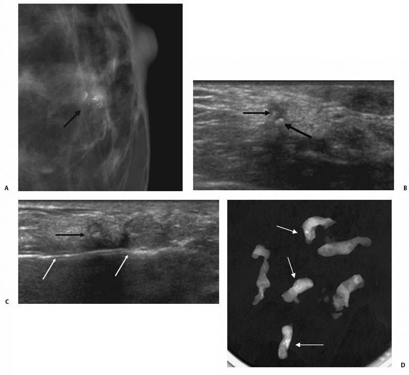
Should typically undergo sampling using stereotactic guidance, but can be identified and undergo biopsy using ultrasound in some cases. In a prospective study, Soo et al identified 23% of 111 lesions on ultrasound that presented as microcalcifications without a mass on mammography, and subsequently performed successful CNB using ultrasound. When calcifications were identified on ultrasound, lesions were more likely to be larger, malignant (69% versus 21%), and invasive when malignancy was diagnosed (72% versus 28%) than lesions not identified on ultrasound. Ultrasound-guidance can be a useful option when percutaneous biopsy using stereotactic guidance is not feasible (Fig. 5.2).


image


Fig. 5.2 A 68-year-old woman with suspicious microcalcifications on mammography who underwent diagnostic ultrasound-guided core needle biopsy. Stereotactic-guided biopsy could not be performed due to small compressed breast thickness. (A) Craniocaudal magnification view of the left breast shows grouped heterogeneous calcifications (arrow). (B) The calcifications were visualized on ultrasound (arrows). (C) Ultrasound-guided vacuum-assisted biopsy was performed by placing the needle (white arrows) posterior to the calcifications (black arrow). (D) Specimen radiograph contains numerous calcifications (arrows). Histology revealed invasive ductal carcinoma and ductal carcinoma in situ.


Ultrasound-Guided Biopsy of Abnormal Axillary Lymph Nodes


  • Can reduce the number of women undergoing sentinel lymph node biopsy.
  • Women with a newly diagnosed invasive breast cancer undergo sentinel lymph node biopsy at the time of lumpectomy or mastectomy (unless there is palpable adenopathy).
  • If FNA of an axillary lymph node in a woman with diagnosed breast cancer shows metastatic breast cancer, she can undergo axillary dissection at the first surgical procedure.
  • If ultrasound shows normal axillary lymph nodes or the ultrasound-guided FNA or CNB is negative, sentinel node biopsy can be performed.

Axillary Adenopathy of Unknown Cause


Contraindications


Contraindications to ultrasound-guided breast biopsy:


  • Uncooperative patient during the procedure

    • Mild sedation using fast-acting oral agents can be helpful if necessary.

  • An uncorrectable known bleeding disorder

    • For these women, FNA or surgical biopsy may be reasonable alternatives.

Women who are not good candidates for stereotactic biopsy due to stroke or small breast size can usually undergo ultrasound-guided breast biopsy if the lesion is identified. The presence of a breast implant can increase the complexity of the approach, but is not necessarily a contraindication.


Preprocedural Evaluation


Equipment


High-Frequency Linear Array Transducers


  • Use of 10 MHz or higher linear transducer is highly recommended.
  • Focal zones should be adjustable rather than fixed in position.

Needle Selection (Core versus Vacuum)


Either automated throw needle (CNB) or vacuum-assisted (VAB) biopsy devices may be used for ultrasound-guided breast biopsy.


Core Needle Biopsy


  • 14-gauge needles are most commonly used for ultrasound-guided CNB, but range in size from 12–18 gauge.
  • Higher diagnostic yields have been demonstrated for 14-gauge needles compared with 16- or 18-gauge core biopsy needles.

Vacuum-assisted Biopsy


  • Needle size ranges from 8–14 gauge.
  • VAB devices may collect single samples that must be retrieved after each pass (similar to CNB devices) or may collect multiple samples with a single needle insertion.
  • VAB is more advantageous for calcified lesions. Bleeding complications may be slightly higher with VAB devices than CNB devices.

VAB can be useful for selective cases:


  • A small lesion that may be difficult to visualize after the first one or two samples. In these cases, the needle can be placed posterior to the lesion and kept stationary during the biopsy. The lesion can be observed for resolution during sampling under real-time.
  • VAB is also useful for the uncommon case when calcifications are sampled by ultrasound rather than stereotactic-guidance as VAB devices that require only one needle insertion often result in less introduction of air that may obscure calcifications.

Fine-Needle Aspiration Biopsy


  • The Radiologic Diagnostic Oncology Group V found the sensitivity and specificity of FNAB of nonpalpable breast lesions to be 85–88% and 56–90%, respectively, which is considerably lower than CNB.
  • FNAB has lower sensitivity for invasive lobular carcinoma compared with invasive ductal carcinoma.
  • CNB should be performed instead of FNAB for most breast lesions.

FNAB can be very useful in


  • The evaluation of cystic lesions of the breast
  • A complicated cyst or complex mass may initially undergo FNAB. If a residual solid component remains, CNB can then be performed.
  • FNAB of suspicious axillary lymph nodes can circumvent the need for sentinel node biopsy in the setting of known breast cancer.

Room Setup


Prior to beginning the procedure, diagnostic breast imaging including any mammograms, ultrasound, and MR images should be reviewed.


  • An examination table that is adjustable in height is convenient to accommodate for radiologist stature.
  • A table that rotates is useful for radiologists who prefer to use their dominant hand to manage the biopsy device.
  • Lights should be dim enough to visualize the lesion well on the ultrasound screen.

Preparation for Procedure


Mar 10, 2016 | Posted by in ULTRASONOGRAPHY | Comments Off on Breast Biopsy

Full access? Get Clinical Tree
Get Clinical Tree app for offline access