and Filiz Özülker1
(1)
Nuclear Medicine, Okmeydani Training and Research Hospital, Istanbul, Turkey
6.1 Case 1: Initial Evaluation of Breast Mass with 18F-FDG PET/CT
History
A 48-year-old female underwent 18F-FDG PET/CT after suspicious mass lesion at left breast was detected on thorax CT and USG.
Findings
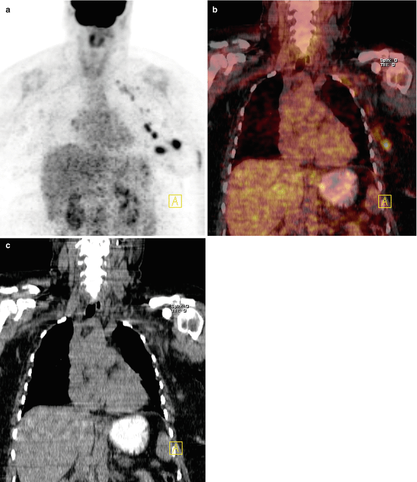
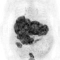
Fig. 6.1
MIP image (a), coronal fusion, and CT images (b, c) show two intensely hypermetabolic foci at lateral aspect of left breast corresponding to mass lesions on CT (SUVmax 9.7). There are also lymph nodes at left axillary fossa and left retroclavicular region showing significant FDG uptake (SUVmax 7.9)
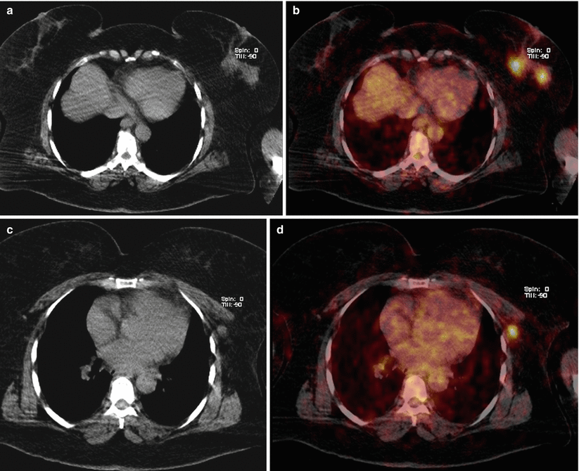
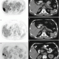
Fig. 6.2
Axial slices of CT and fusion images (a–d) show mass lesions at left breast and lymph nodes at left axillary fossa which show increased FDG uptake
Interpretation
Intense FDG uptake at left breast mass lesions is indicative of malignant disease of breast and hypermetabolic foci at left axilla and retroclavicular region reflect metastatically involved lymph nodes.
Result
Biopsy of the mass lesion at left breast turned out to be invasive ductal carcinoma.
Teaching Point
18F-FDG PET/CT has high specificity in the detection of metastatic axillary lymph nodes, but sensitivity is not satisfactory especially in lymph nodes with low tumor burden. So 18F-FDG PET/CT may preclude axillary dissection with sentinel lymph node mapping when FDG positive lymph nodes are detected.
6.2 Case 2: Invasive Ductal Carcinoma and Fibroadenoma
History
A 26-year-old female status post biopsy proven to be invasive ductal carcinoma at right breast and fibroadenoma at left breast underwent 18F-FDG PET/CT for pretreatment staging.
Findings
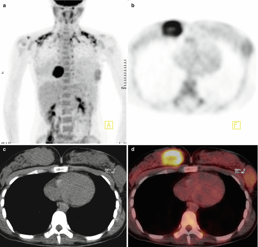
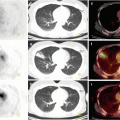
Fig. 6.3
MIP image (a), axial slices of PET, CT, and fusion images (b, c, d) show intensely hypermetabolic round mass lesion at right breast measuring 5.7 × 5 cm and having hypometabolic necrotic center (SUVmax 10). There is also another mass lesion located laterally at left breast measuring 4 × 4 cm and showing mild FDG uptake (SUVmax 3). Increased FDG uptake at supraclavicular and paravertebral areas representing uptake at brown adipose tissue (BAT) is also noted
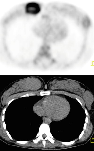
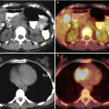
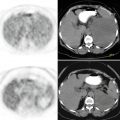
Fig. 6.4
Axial PET and CT images
Interpretation
Intense FDG uptake at right breast mass lesion is indicative of a malignant disease, whereas mild uptake at left breast mass lesion is suggestive for a benign lesion but malignancy cannot be ruled out.
Result
Histopathology revealed invasive ductal carcinoma at right breast and fibroadenoma at left breast.
Teaching Point
18F-FDG PET/CT has good sensitivity in detecting invasive ductal carcinomas. Benign neoplasms like ductal adenoma, fibrous dysplasia, and fibroadenomas may give false positive results and should be taken into account while evaluating breast lesions. Low levels of SUVmax at benign lesions might help to discriminate them from malignancies.
6.3 Case 3: Pretreatment Staging in Locally Advanced Breast Cancer
History
A 52-year-old female with right sided locally advanced breast cancer (LABC) (invasive ductal carcinoma) underwent 18F-FDG PET/CT for pretreatment staging.
Findings
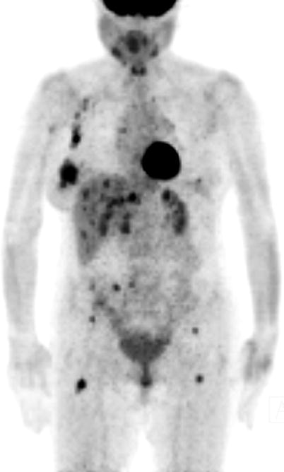
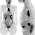
Fig. 6.5


MIP image shows intensely hypermetabolic lesion at right breast, hypermetabolic right axillary lymph nodes, multiple hypermetabolic foci at liver, hypermetabolic lesions at pelvic bones and both proximal femurs
Related posts:
Stay updated, free articles. Join our Telegram channel
Full access? Get Clinical Tree
