and Filiz Özülker1
(1)
Nuclear Medicine, Okmeydani Training and Research Hospital, Istanbul, Turkey
10.1 Case 1: Staging of Hepatocellular Carcinoma
History
A 66-year-old male underwent 18F-FDG PET/CT after having diagnosed as hepatocellular carcinoma (HCC).
Findings
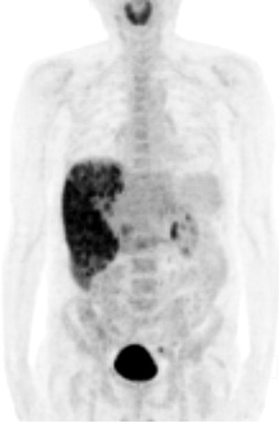
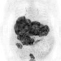
Fig. 10.1
MIP image shows heterogenously increased FDG uptake at liver. There is not any other pathological FDG uptake detected at rest of the body
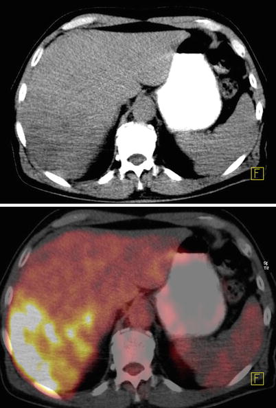
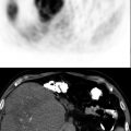
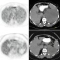
Fig. 10.2
Axial CT and fusion images show heterogenous hypermetabolism at right lobe of liver
Interpretation
Hypermetabolism at liver is compatible with primary hepatic malignancy.
Teaching Point
HCC exhibit variations in FDG accumulation for unknown reasons. Low or absent FDG uptake can be seen in low-grade hepatocarcinoma, and this fact is attributed to the presence of a higher glucose-6-phosphatase activity resulting in a low FDG uptake.18F-FDG PET CT may give additional information in the routine staging as a complementary imaging tool to dynamic CT of liver and chest x-ray.
10.2 Case 2: Restaging of Hepatocelular Carcinoma
History
A 63-year-old male who had undergone hepatic resection for HCC 1 year ago underwent 18F-FDG PET/CT for restaging.
Findings
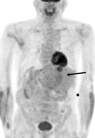
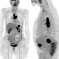
Fig. 10.3


MIP image shows focal FDG uptake at liver. Displacement of liver to left side of abdomen is also noted (arrow)
Related posts:
Stay updated, free articles. Join our Telegram channel
Full access? Get Clinical Tree
