and Filiz Özülker1
(1)
Nuclear Medicine, Okmeydani Training and Research Hospital, Istanbul, Turkey
13.1 Case 1: Surrenal Adenoma
History
A 52-year-old male without any known primary malignancy underwent 18F-FDG PET/CT after a left adrenal incidentaloma was detected by abdominal MRI.
Findings
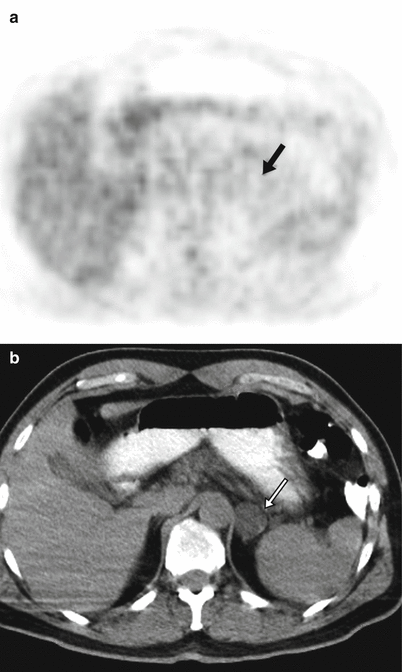
Fig. 13.1
Axial slices of CT and PET (a, b) show homogeneous, well-defined, 5.4-HU round mass with dimensions of 4.5 × 4 × 3.5 cm in the left adrenal gland without any significant FDG uptake (arrow). MRI (not shown here) displayed a drop in signal intensity on out of phase imaging
Interpretation
This finding is diagnostic of a benign adrenal incidentaloma.
Teaching Point
Adrenal adenomas are neoplasms which are either functional or not and incidentally detected on conventional anatomic imaging. They are typically less than 3 cm in diameter, well marginated, and of uniform and less than 10 HU attenuation at unenhanced CT. Approximately 30 % of adenomas are lipid poor with an attenuation greater than 10 HU. They show marked contrast material washout at delayed imaging at contrast enhanced CT and drop in signal intensity on out of phase chemical shift MR imaging.
13.2 Case 2: Surrenal Adenoma
History
A 55-year-old male without any known primary malignancy underwent 18F-FDG PET/CT for evaluation of a left adrenal incidentaloma.
Findings
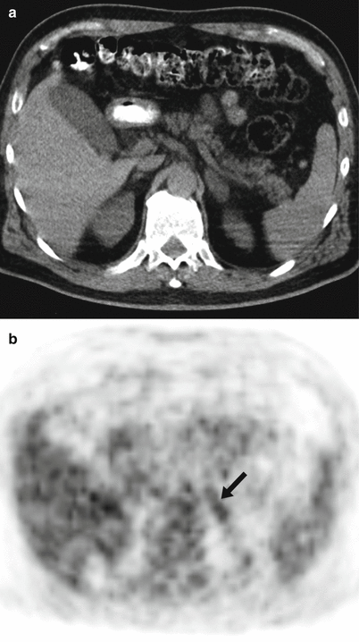
Fig. 13.2
Axial slices of CT and PET (a, b) show mildly increased FDG uptake at left oval surrenal mass measuring 2 × 2 cm (SUVmax 3) (arrow). SUV ratio (nodule SUV(max)/liver SUV(avg)) is 3/2.7 = 1.1. MRI (not shown here) displayed a drop in signal intensity on out of phase imaging. The lesion had a mean HU of −13 on CT images
Interpretation
This finding is indicative of likelihood for a benign adrenal incidentaloma.
Teaching Point
It has been proposed that identification of malignancy at surrenal gland can be accomplished by applying an SUVmax cutoff of greater than 3.1 and SUV ratio of greater than 1.0. Combined PET/CT with mean attenuation >10 HU and SUV(max) >3.1 had 97.3 % sensitivity and 86.2 % specificity in identification of malignancy, and combined PET/CT with mean attenuation >10 HU and SUV ratio >1.0 had 97.3 % sensitivity and 74.1 % specificity.
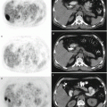
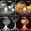
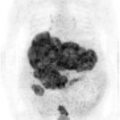
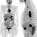
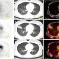
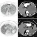
13.3 Case 3: Adrenal Schwannoma
History
A 32-year-old woman was incidentally found to have a left adrenal mass of 4 × 8 × 10 cm on abdominal ultrasonography. The patient underwent 18F-FDG PET/CT and then left adrenalectomy was performed.
Findings
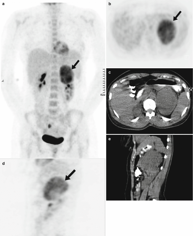
Fig. 13.3
MIP (a), axial (b, c), and sagittal (d, e) PET and CT images show intense uptake within the left adrenal mass (arrows)
Interpretation
Metabolic activity of the lesion is at the level of malignancy, and when the size and the imaging characteristics of the lesion is taken into account, the lesion is strongly suspicious of a malignant adrenal neoplasm.
Result
Histological examination revealed the diagnosis of schwannoma. This diagnosis was supported by immunohistochemistry of S-100 and vimentin positivity.
Teaching Point
Although adrenal lesions with a SUVmax >3 are thought to be malignant, benign lesions like schwannoma can cause false positive results.
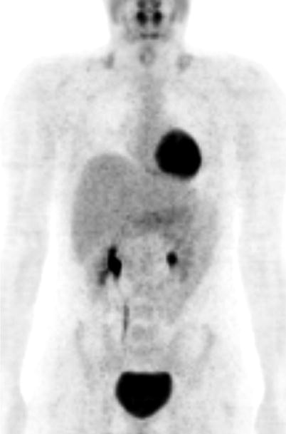
13.4 Case 4: Adrenal Ganglioneuroma
History
A 18-year-old male patient underwent CT, which showed a relatively homogenous left adrenal tumor measuring 5.2 × 4.3 × 7.1 cm. 18F-FDG PET/CT scan was performed to rule out malignancy at primary lesion and detect any possible metastases.
Findings
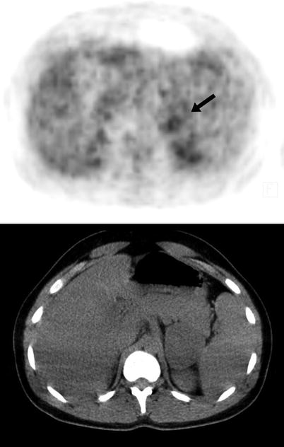
Fig. 13.4
Axial slices of PET and CT images show mild uptake at left adrenal mass (arrow) (SUVmax 4.1)
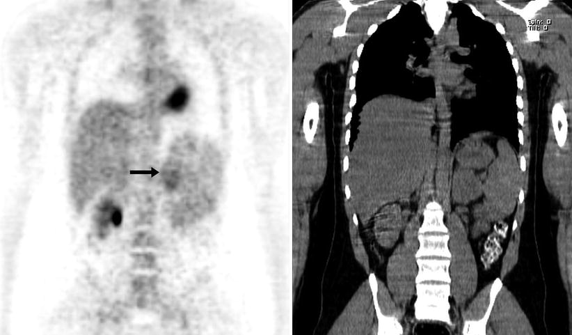
Fig. 13.5
Coronal PET and CT images show mass lesion at left adrenal gland with subtle FDG uptake (arrow)