and Filiz Özülker1
(1)
Nuclear Medicine, Okmeydani Training and Research Hospital, Istanbul, Turkey
17.1 Case 1: Basal Cell Carcinoma
History
A 84-year-old female status postsurgical excision of a lesion from skin of nose proven to be basal cell carcinoma underwent 18F-FDG PET/CT scan.
Findings
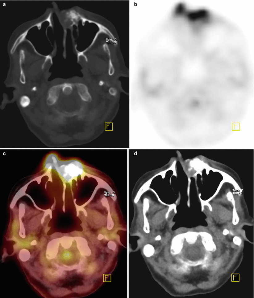
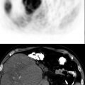
Fig. 17.1
Axial slices of CT, PET, and fusion (a–d) show significantly increased FDG uptake at left side of dorsum of nose (SUVmax 5.9)
Interpretation
This finding is compatible with local recurrence of the disease.
Teaching Point
Although there are limited data, 18F-FDG PET/CT imaging might be able to image and identify basal cell carcinoma in the head and neck region. The histologic subtype of the basal cell carcinoma appears to affect the ability of detection with FDG PET, with the nodular histologic subtype being more FDG avid.
17.2 Case 2: Squamous Cell Carcinoma of Skin
History
A 77-year-old female who had been diagnosed to have squamous cell carcinoma of nasal skin following surgical excision, underwent 18F-FDG PET/CT.
Findings
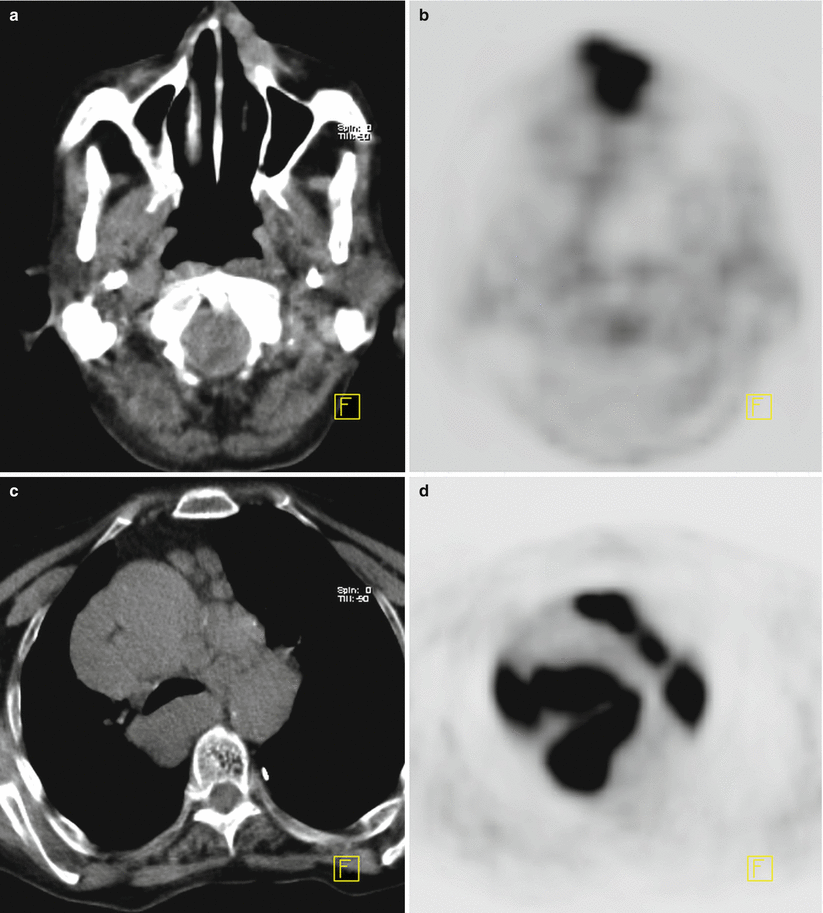
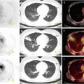
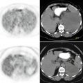
Fig. 17.2
Axial slices of CT and PET images of cranium (a, b) and thorax (c, d) show intensely increased FDG uptake at left side of dorsum of nose (SUVmax 8.7), multiple intensely hypermetabolic lymph nodes at mediastinum (SUVmax 16)
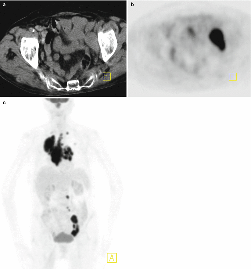
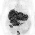
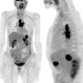
Fig. 17.3
Axial slices of CT and PET (a, b) and MIP (c) images show intensely hypermetabolic lymph nodes at left obturator area (arrow) and mediastinal and intraabdominal lymph node stations (SUVmax 16)
Interpretation
Hypermetabolic lesion at left side of nose is compatible with local recurrence of the disease. Hypermetabolic lymph nodes are suspicious for lymphoproliferative malignant disease.
Result
Biopsy from mediastinal lymph nodes revealed granulomatous disease.
Teaching Point
18F-FDG PET CT might be a sensitive tool in detecting local recurrence in cutaneous squamous cell carcinoma but lack of specificity may cause false positive results in detection of lymph node metastases.
Granulamatous diseases should always be kept in mind when multiple hypermetabolic lymph nodes are seen.
17.3 Case 3: Malignant Degeneration of a Burn Scar into a Squamous Cell Carcinoma
History
A 36-year-old male had an electrical burn at right foot approximately 1 year ago. Biopsy from the exophytic lesion developed at chronic burn scar revealed squamous cell carcinoma. The patient underwent 18F-FDG PET/CT scan for staging.
Related posts:
Stay updated, free articles. Join our Telegram channel

Full access? Get Clinical Tree


