Breast Cancer, an Overview
Breast cancer is a common, life-threatening disease. If skin cancers are excluded, breast cancer is the most frequently diagnosed malignancy and a leading cause of cancer mortality among women, second only to lung cancer (1). Women have a 12.5% (1 in 8) lifetime risk for developing breast cancer by 85 years of age (2,3). Although breast cancer incidence rates increased about 4% per year in the 1980s, they have leveled off at about 110 cases per 100,000 women (2,3).
Women have a 12.5% (1 in 8) lifetime risk for developing breast cancer by 85 years of age.
The National Cancer Institute’s Surveillance, Epidemiology, and End Results (SEER) program reported decreases in the breast cancer mortality rate for 1989 to 1992 across all age groups. An 8.1% decrease in the mortality rate was reported for 40- to 49-year-old women compared with a 9.3% decrease for 50- to 59-year-old women, a 4.8% decrease for 60- to 69-year-old women, and 3.1% decrease for 70- to 79-year-old women (4). It is estimated that 41,200 breast cancer-related deaths will occur in 2003 (40,800 in women, 400 in men) compared with 43,884 deaths in 1995 when the number of deaths was the highest (1). These decreases in mortality are at least partially attributable to the benefits gained by early detection through screening mammography.
It is estimated that 211,300 women and 1,300 men will be diagnosed with invasive ductal carcinoma and that 41,200 (40,800 women and 400 men) breast cancer-related deaths will occur in 2003.
RISK FACTORS
Many factors are reportedly associated with an increased risk for breast cancer, but only a few are considered significant. Included among these are gender, age, a personal or family history of breast cancer, prior breast biopsy with certain histologic diagnoses, mutations in the BRCA1 and BRCA2 genes, and several other relatively rare breast cancer susceptibility genes. Other factors that have been implicated include early menarche, late menopause, late first-term pregnancy, nulliparity, and postmenopausal obesity. For these risk factors, prolonged exposure of breast tissue to estrogen is thought to play a role. A history of high-dose radiation therapy to the chest is another risk factor for breast cancer (1,2).
Significant risk factors for breast cancer include gender, age, personal history of breast cancer, first-degree family member with breast cancer, prior breast biopsy with a risk marker lesion, and mutations in breast cancer susceptibility genes.
Women are more likely to develop breast cancer. It is estimated that 211,300 women will be diagnosed with invasive ductal carcinoma in 2003 compared with 1,300 men. The incidence of breast cancer increases with advancing patient age. The risk increases rapidly in premenopausal women and more slowly in postmenopausal women. Women with a
personal history of breast cancer are at a higher risk for developing another breast cancer compared with women who have never had breast cancer. Women with one or multiple first-degree relatives with breast cancer (mother, father, sisters, brothers, daughters, and sons) are at increased risk, particularly if the breast cancers developed premenopausally are bilateral, or are multiple. Women who have had a breast biopsy, particularly if atypical ductal hyperplasia (ADH) or lobular neoplasia (lobular carcinoma in situ, LCIS) was diagnosed, are also at increased risk. With ADH, the lesion itself may represent a precursor, in contrast to lobular neoplasia, which is considered to be more of a marker lesion. Two genes (BRCA1 and BRCA2) have been isolated in some women with breast cancer. It is estimated that 5% to 10% of all breast cancers in the United States are hereditary, related to mutations in the BRCA1 or BRCA2 genes. Women with mutations in the BRCA1 gene are also at increased risk for developing ovarian cancer. The risk for developing breast cancer in women who carry mutations in this gene is 85% by 70 years of age (63% by 70 years of age for ovarian cancer). Women who carry mutations in the BRCA2 gene are at increased risk for developing breast cancer but not ovarian cancer. Hereditary male breast cancer is associated with mutations in the BRCA2 gene but not the BRCA1 gene. Some genetic diseases (e.g., Li-Fraumeni syndrome, Cowden’s disease, and ataxia-telangiectasia) are associated with breast cancer susceptibility genes and with an increased risk for breast cancer (2,3,5).
personal history of breast cancer are at a higher risk for developing another breast cancer compared with women who have never had breast cancer. Women with one or multiple first-degree relatives with breast cancer (mother, father, sisters, brothers, daughters, and sons) are at increased risk, particularly if the breast cancers developed premenopausally are bilateral, or are multiple. Women who have had a breast biopsy, particularly if atypical ductal hyperplasia (ADH) or lobular neoplasia (lobular carcinoma in situ, LCIS) was diagnosed, are also at increased risk. With ADH, the lesion itself may represent a precursor, in contrast to lobular neoplasia, which is considered to be more of a marker lesion. Two genes (BRCA1 and BRCA2) have been isolated in some women with breast cancer. It is estimated that 5% to 10% of all breast cancers in the United States are hereditary, related to mutations in the BRCA1 or BRCA2 genes. Women with mutations in the BRCA1 gene are also at increased risk for developing ovarian cancer. The risk for developing breast cancer in women who carry mutations in this gene is 85% by 70 years of age (63% by 70 years of age for ovarian cancer). Women who carry mutations in the BRCA2 gene are at increased risk for developing breast cancer but not ovarian cancer. Hereditary male breast cancer is associated with mutations in the BRCA2 gene but not the BRCA1 gene. Some genetic diseases (e.g., Li-Fraumeni syndrome, Cowden’s disease, and ataxia-telangiectasia) are associated with breast cancer susceptibility genes and with an increased risk for breast cancer (2,3,5).
A diagnosis of atypical ductal hyperplasia or lobular neoplasia (lobular carcinoma in situ) on a prior breast biopsy is associated with a significant risk for the subsequent development of breast cancer.
Early menarche and the establishment of regular menses are risk factors in breast cancer development. It is estimated that the risk for breast cancer decreases by 10% to 15% with each year or two that menarche is delayed (2,3). Late menopause is another factor associated with increased breast cancer risk. The risk for breast cancer can be decreased by 50% with bilateral oophorectomy before 40 years of age, and it is estimated that women undergoing natural menopause before 45 years of age have half the risk for developing breast cancer than women who undergo menopause after 55 years of age (5). Breast cancer risk is reduced by 50% if the first term pregnancy occurs before 20 years of age compared with after 35 years of age (2,3,5). Women who do not have any pregnancies (nulliparous) and those who are obese after menopause are at increased risk compared with women who have had children and those who are not obese after menopause. Patients who undergo high-dose radiation therapy to the chest for lymphoma or an enlarged thymus are at increased risk for breast cancer (2,3,5). Breast cancer in these patients develops about 10 to 15 years after the radiation therapy and tends to be aggressive.
About 5% to 10% of all breast cancers are estimated to be related to mutations in the BRCA1 or BRCA2 genes.
Women with mutations in the BRCA1 gene are at increased risk for developing breast and ovarian cancer.
Hereditary male breast cancer is associated with mutations in BRCA2.
Other factors reportedly associated with increased breast cancer risk are controversial. These include oral contraceptive use, alcohol intake, exogenous estrogen use (after 10 years of use), and dietary fat intake (saturated fats). Lactation, exercise, and monounsaturated fat intake may have protective benefits. Although implicated in some studies as a risk factor, cigarette smoking does not appear to be associated with an increased risk for breast cancer (2,3,5).
Patients with a history of high-dose radiation to the chest wall have an increased risk for developing breast cancer 10 to 15 years after the radiation.
SCREENING MAMMOGRAPHY AND SCREENING RECOMMENDATIONS
The use and benefits of screening mammography in diagnosing breast cancer have been controversial. The goal of screening mammography is to identify breast cancer as early as possible before it becomes apparent clinically as a lump or with skin changes or distant metastasis. But, how do we know that mammography can show early breast cancers consistently? And if it can, how do we know that finding these early breast cancers is of any benefit to the patient? The ability of mammography to demonstrate small breast cancers consistently and the benefits of detecting these breast cancers through mammography were established by several randomized controlled trials.
Specifically, seven randomized controlled trials of screening mammography have all proved a benefit. Fewer women die from breast cancer among the population invited to
undergo screening mammography compared with the control group of women not invited to undergo screening mammography. The reported benefits range from a 20% to 40% reduction in breast cancer mortality among the women invited to screening mammography (6, 7, 8). Screening mammography does show breast cancers consistently, and the diagnosis and treatment of the women with breast cancers identified through mammography saves lives.
undergo screening mammography compared with the control group of women not invited to undergo screening mammography. The reported benefits range from a 20% to 40% reduction in breast cancer mortality among the women invited to screening mammography (6, 7, 8). Screening mammography does show breast cancers consistently, and the diagnosis and treatment of the women with breast cancers identified through mammography saves lives.
Several randomized controlled trials have been done to establish the ability of mammography to demonstrate small breast cancers leading to significant reductions in breast cancer mortality rates.
The issue of screening women who are 40 to 49 years of age has been particularly controversial. However, a statistically significant benefit has been reported from several of the randomized controlled trials, including the Gothenberg and Malmo trials, which reported 44% and 36% reductions in breast cancer mortality, respectively, in this population of women. Also, a statistically significant benefit (26%) of screening 40- to 49-year-old women was reported from a metaanalysis (a statistical test that combines data reported from multiple trials) that used data from the seven population-based screening trials (9). In determining appropriate screening intervals, it is important to consider tumor sojourn times. Tumor sojourn time, defined as the time taken for cancers to go from preclinical to clinical mammographic detectability, is 1.7 years in premenopausal women and 3.3 years in postmenopausal women (10). Screening women in their 40s, therefore, is optimally done at annual intervals.
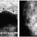
Mammography is not a perfect test. In some women with breast cancer, the cancer is not visible on the mammogram, even under the best of circumstances. The false-negative rates for mammography range from 7% to 15% (11). This is why it is important that women recognize the need for breast self-examination and annual breast examinations by a health care provider. Although there are false-negative mammograms, screening mammography is an effective method for finding early, potentially curable breast cancers and saving lives. It is the best and, currently, the only, reliable defense women have against breast cancer.
Reported false-negative rates for mammography range from 7% to 15%. False-negative rates vary depending on breast tissue density and lesion type (higher false-negative rates are reported with invasive lobular carcinoma).
Using the cumulative data from all of the randomized controlled screening trials, the American Cancer Society (ACS), in March 1997, issued new screening guidelines recommending annual screening mammography for all women starting at 40 years of age. The baseline study (35 to 40 years of age) is no longer part of the screening guidelines. The ACS also recommends monthly breast self-examination starting at 20 years of age and an annual physical examination by a health care provider every 3 years for women 20 to 33 years of age and annually starting at 40 years of age. Breast cancer is now more commonly diagnosed after screening mammography compared with previously, when most breast cancers were diagnosed by the patient during breast self-examination (12).
Mammography is recommended annually starting at 40 years of age for women at average risk for breast cancer.
Although there have been no rigorous trials to evaluate the use and benefit of screening mammography in high-risk women, many radiologists recommend starting screening mammography at 30 years of age in women in a high-risk category, including those with a family history of breast cancer (first-degree relative), particularly if the family member developed premenopausal, bilateral, or multiple breast cancers. In a woman whose mother developed breast cancer before 40 years of age, consider starting mammographic screening 10 years before the age of detection in the mother. For example, if the mother developed breast cancer at 36 years of age, consider starting mammographic screening in the daughter at 26 years of age.
At this time, there are insufficient data to establish screening guidelines for women with an increased risk for breast cancer. Options to consider in these women include starting annual screening mammography at 30 years of age, employing shorter screening intervals (e.g., every 6 months), or adding ultrasound or magnetic resonance imaging to the screening examination.
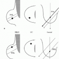
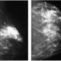
Mammography provides the ability to diagnose stage 1 and 2 breast cancers as well as, in many patients, noninvasive, intraductal (stage 0) breast cancers. The diagnosis of early, potentially curable breast cancer increases the treatment options for patients and probably renders available treatments more effective. Regardless of the histologic grade (aggressiveness) of a tumor or the status of the axillary lymph nodes, women with breast cancers that are smaller than 1 cm in size have a 12-year survival rate of 95% (13). The goal of screening mammography, therefore, becomes the identification of cancers that are smaller than 1 cm in size. Inclusion of all breast tissue in the images through optimal mammographic positioning; high-contrast, high-resolution, well-exposed images; and the interpretive skills of the radiologist become pivotal in our ability to identify cancers consistently that are less than 1 cm in size.
MAMMOGRAPHY QUALITY STANDARDS ACT
Stay updated, free articles. Join our Telegram channel

Full access? Get Clinical Tree


