Box 4.1 Key Mammographic Signs of Breast Cancer
- Ill-defined, irregular or spiculated mass or masses
- Pleomorphic microcalcifications, often with a linear or ductal distribution and with or without an associated mass or soft tissue density
- Associated features include:
- architectural distortion
- skin thickening
- axillary lymph node enlargement
- architectural distortion
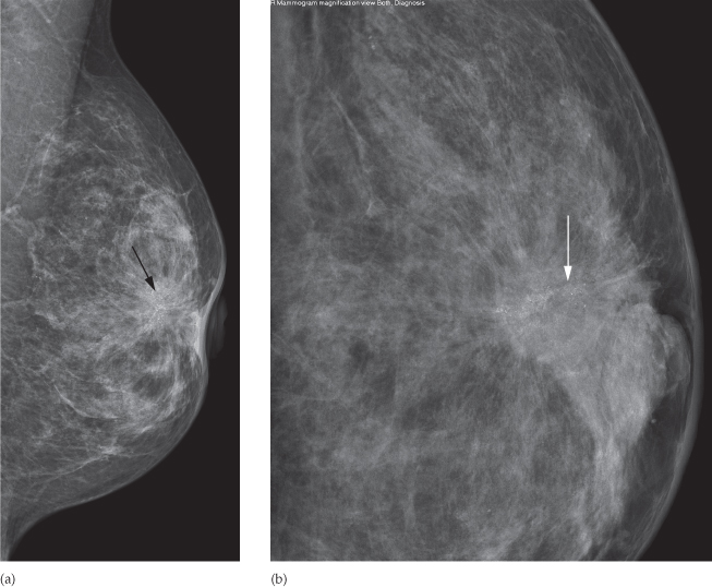
Fig. 4.1 Mammographic images of breast cancer. (a) Left mediolateral oblique and (b) compressed magnified left craniocaudal views of an irregular spiculated mass (black arrow) with associated pleomorphic microcalcification (white arrow) behind the nipple with associated architectural distortion and nipple retraction.
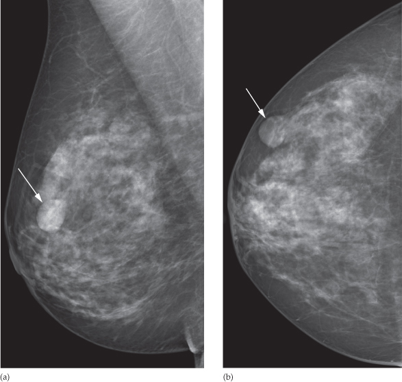
Benign masses are usually well defined and ovoid or spherical (Fig. 4.2). They may contain calcifications, but these are generally large and coarse in appearance.
Fig. 4.2 Mammographic image of a benign breast lesion. (a) Right mediolateral oblique and (b) craniocaudal views of the right breast demonstrating a very well-defined ovoid mass in the upper outer quadrant of the breast (arrow).
Breast Ultrasound
Related posts:
Stay updated, free articles. Join our Telegram channel

Full access? Get Clinical Tree


