Cancer Biology


Cancer Biology


Tissue homeostasis depends on the regulated cell division and self-elimination (programmed cell death) of each of its constituent members except its stem cells. In fact, a tumor arises because of uncontrolled cell division and failure for self-elimination. One can consider cancer as a Darwinian-like process whereby the fittest cells reproduce to become the dominant population of a tumor. Alterations in three groups of genes are responsible for the deregulated control mechanisms that are the hallmarks of cancer cells:


  • Proto-oncogenes are components of signaling networks that act as positive growth regulators in response to mitogens, cytokines, and cell-to-cell contact. A gain-of-function mutation in only one copy of a proto-oncogene results in a dominantly acting oncogene that often fails to respond to extracellular signals.


  • Tumor suppressor genes are also components of the same signaling networks as protooncogenes, except that they act as negative growth regulators. They modulate proliferation and survival by antagonizing the biochemical functions of proto-oncogenes or responding to unchecked growth signals. In contrast to oncogenes, inactivation of both copies of tumor suppressor genes is required for loss of function in most cases.


  • DNA stability genes form a class of genes involved in both monitoring and maintaining the integrity of DNA. Loss of these genes results in defective sensing of DNA lesions as well as improper repair of the damaged template.

The malignant progression from normal tissue to tumor to metastasis occurs in various “steps” over a period of time. These steps, which are the result of mutations, deletions,
or gene changes in the three groups of genes described previously, may occur spontaneously because of random errors or result from exposure to agents as diverse as chemical mutagens, ionizing radiations, ultraviolet light, and viruses; and provide a growth or survival advantage that allows the cells to become the clonal origin of the tumor. To summarize, tumor evolution results from the accumulation of gene mutations that arise in a single cell that has suffered a disruption in its regulatory mechanisms for proliferation, self-elimination, immortalization, and genetic stability. This is illustrated in Figure 18.1.


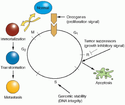
FIGURE 18.1 The process of malignant transformation results from mutations in three groups of genes: gain-of-function mutations that activate oncogenes, loss-of-function mutations that inactivate tumor suppressor genes, and loss of activity of DNA stability (e.g., repair) genes that increases the probability for genomic instability. This figure depicts how the stimulatory effects of oncogenes on the cell cycle are opposed by the inhibitory effects of tumor suppressor genes on the cell cycle that can lead to apoptosis. R indicates the restriction point that is regulated by the p53 and pRb tumor suppressor genes. The consequences of oncogene activation and tumor suppressor gene and DNA integrity gene inactivation are immortalization, transformation, and metastasis.


▪ MECHANISMS OF CARCINOGENESIS

A single genetic alteration that leads to the activation of an oncogene or loss of a tumor suppressor gene does not, by itself, lead to the formation of a solid tumor. Instead, carcinogenesis appears to be a multistep process with multiple genetic alterations occurring over an extended period of time; at least, that is how it appears. Sometimes, these genetic alterations are carried in the germ line, like, for example, in the cancer- predisposing syndrome retinoblastoma; however, heritable mutations are rare. Most alterations that lead to cancer are acquired in the form of somatic mutations: chromosomal translocations, deletions, inversions, amplifications, or simple point mutations.

Initially, it was thought that cancer was the result of deregulated growth signals by oncogenes, a concept supported by increased proliferation in many types of cancer. In the last decade, the finding that many cancers possess diminished apoptotic (programmed cell death) programs or loss of cell cycle control has led to the concept that mutations in proto-oncogenes and tumor suppressor genes that inhibit apoptosis provide a selective growth advantage to a premalignant cell that allows it to clonally expand. Mutations in DNA stability genes increase the rate of acquiring genetic mutations that will result in a malignant tumor. Thus, although tumor cells are considered clonal in origin, most tumors contain heterogeneous populations of cells that differ in their ability to repopulate the tumor or form metastases. In fact, only a small percentage of tumor cells possess the ability to form a tumor, leading to the concept that tumors possess “stem cells” and that elimination of these stem cells is essential for controlling tumor growth.


▪ ONCOGENES

The first demonstration that a tumor was initiated by a cellular component found in tumor cells but not in normal cells was shown by Rous in the early 1900s. His landmark studies demonstrated that cell-free extracts derived from chicken sarcomas could cause a new sarcoma, if injected into healthy chickens. In the 1970s,
with the advent of molecular biology, several groups identified the etiological agent for sarcoma formation in chickens as an RNA virus, designated as the Rous sarcoma virus (RSV), which belongs to a group of viruses designated as retroviruses—viruses whose genomes are composed of RNA. Thus, oncogenes were first discovered from a study of retroviruses that cause cancers in animals. Although the virus had been identified, it still remained to be elucidated how this retrovirus causes a sarcoma because another virus belonging to this same group of RNA viruses, avian leukosis virus (ALV), does not transform cells in culture or induce sarcomas. Analysis of the genomes of ALV and RSV revealed that RSV contains approximately 1,500 more base pairs (bp) of DNA than ALV. It was hypothesized, therefore, that these extra base pairs of DNA in the RSV genome are responsible for the tumorigenic activity. This was supported by an observation that deletion mutants of RSV that are missing this 1,500-bp region lose their transformation potential, but can still replicate and produce viral progeny normally. This led to the conclusion that the transforming activity and replicative activity of RSV are encoded by genetically distinct regions of the virus and that only a small portion of the RSV genome is needed for transformation.


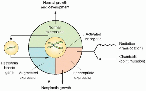
FIGURE 18.2 How the concept of oncogenes provides a ready answer for how agents as diverse as viruses, radiations, and chemicals all can induce tumors that are essentially indistinguishable one from another. The retrovirus inserts a gene; a chemical may activate an endogenous oncogene by a point mutation; radiation may do the same by causing, for example, a translocation. (Adapted from Bishop JM. Cellular oncogene retroviruses. Ann Rev Biochem. 1983;52:301-354, with permission.)

From these early studies, several important conclusions could be derived. These are the following:


  • Cancer can be caused by a genetically transmissible agent—in the case of chicken sarcoma, by a retrovirus containing a unique piece of genetic information that was later designated as the src gene;


  • Only a certain region of a retrovirus is needed for transformation; and


  • The region of the viral genome necessary for transformation is not involved in the normal replicative life cycle.

Huebner and Todaro later proposed that cancercausing viral genes such as src are normally inactive but can be activated when they recombine with a retroviral genome. Once they do so, they pass from being a benign proto-oncogene (i.e., c-src) to a malignant form (v-src) capable of causing cancer when introduced into the appropriate host cell. Although we now know that viruses represent only one of several mechanisms that cause the deregulated expression of a proto-oncogene, these studies helped to define oncogenes as mutant forms of normal cellular genes that are altered in expression and/or function by various agents, including radiation, chemicals, and viruses. Consequently, very different agents produce tumors that are indistinguishable from one another. This is illustrated in Figure 18.2.


▪ MECHANISMS OF ONCOGENE ACTIVATION

Although many mechanisms are involved in oncogene activation, transcriptional deregulation by overexpression or abnormal expression of the messenger RNA(mRNA) of a proto-oncogene is a common theme. At least four mechanisms exist for oncogene activation in human neoplasms (Fig. 18.3).


Retroviral Integration through Recombination

Retroviral integration of proto-oncogene sequences in retroviral genomes occurs through two possible recombination mechanisms. First, mRNAs from a proto-oncogene recombine with viral genomic RNAs. During the recombination process, the proto-oncogene mRNA becomes deregulated as it comes under the control of the viral promoter, termed long terminal repeat (LTR). However, the probability of RNA recombination events between proto-oncogene mRNA and viral mRNA generating an oncogenic retrovirus is quite low and undermines the importance of this mechanism.


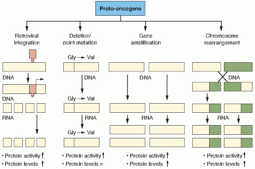
FIGURE 18.3 Four basic mechanisms of how a proto-oncogene can become an activated oncogene. Retroviral integration in proximity to a proto-oncogene results in transcriptional control of the proto-oncogene by the viral promoter, resulting in increased proto-oncogene protein and activity. Deletion or point mutations in the proto-oncogene result in increased activity of the proto-oncogene without necessarily changing transcription or protein levels of the proto-oncogene. Increased copy number of a proto-oncogene by gene amplification results in increased transcription, protein levels, and activity of the proto-oncogene. Chromosome translocation results in an altered proto-oncogene product that can have increased transcription, protein levels, and activity. All of these alterations in a proto-oncogene that occur at the DNA level manifest themselves at the protein level as increased activity and, in some cases, increased protein levels.

A second more probable mechanism is as follows: First, a retroviral genome integrates in proximity to a proto-oncogene, where the proto-oncogene is under the transcriptional control of the retrovirus LTR promoter. Then the viral and proto-oncogene sequences become closely associated through a DNA recombination event that permits the production of mRNAs that contain both viral and proto-oncogene sequences. In this scenario, the proto-oncogene becomes transcriptionally deregulated because it is under the control of the viral promoter LTR. In addition, it can acquire mutations in its coding sequence. Although the proto-oncogene can become mutated during the recombination process, the key point is that its
deregulated expression by the viral LTR increases its expression and promotes cell growth.


DNA Mutation of Regulatory Sites

The union of the technique for gene transfer with mouse transformation assays facilitated the isolation of human oncogenes that were activated by DNA mutation. Transfection of human DNA into immortalized but untransformed mouse cells was first used to isolate the H-ras oncogene from bladder carcinoma cells. The key to this approach is that only transformed cells possess the ability to grow in soft agar (Fig. 18.4). The implicit assumption is that a specific gene (or more) is responsible for causing the bladder carcinoma, and that it will act in a dominant fashion to induce a tumor. Indeed, multiple groups were successful in isolating the H-ras oncogene by this approach.


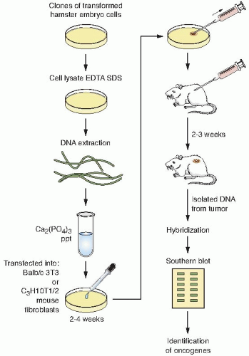
FIGURE 18.4 Schematic diagram of a typical DNA transfection protocol in which oncogenes can be isolated from cells transformed in vitro by either radiation or chemical carcinogens. DNA sequences are then characterized using Southern blot hybridization.

Several steps are needed for the molecular cloning of an oncogene from transformed rodent cells using the rodent fibroblast transformation assay. These are the following:


  • The human DNA containing the transforming oncogene is transfected into mouse cells.


  • The DNA from the transformed mouse cells is serially transfected to reduce the amount of human DNA that is not associated with the transforming oncogene.


  • After several rounds of transfection, the DNA is isolated from a soft agar colony and digested with restriction enzymes to make a genomic DNA library.


  • The library is then screened with a human-specific repetitive probe that does not crossreact with the mouse’s DNA, thereby identifying human sequences in a mouse background.


  • Clones that possess human repetitive sequences are then isolated and digested with restriction enzymes to identify a similar-length fragment that is common to all transformants.


  • Finally, the DNA from the clones is transfected into mouse cells to confirm its oncogenic potential. If the oncogene is present in this genomic clone, then a significant percentage of the transfected mouse cells should be transformed when compared with transfecting genomic DNA from untransformed cells.

Perhaps the prototypical example of oncogene activation by DNA mutation is the H-ras oncogene. The H-ras oncogene was isolated by the approach recently described, and its DNA sequence was compared with its normal cellular counterpart. At first comparison, there did not seem to be any difference between oncogenic and proto-oncogenic forms. However, because H-ras is a relatively small oncogene, it was possible to sequence the entire gene to rigorously search for small mutations. It did not take long to find the difference between the two forms of the gene. The transforming oncogenic H-ras gene possesses a single bp mutation that changes the 12th amino acid from glycine to valine. This single DNA mutation is responsible for changing H-ras from a benign proto-oncogene into a malignant oncogene. We now know that mutations in codons 13 and 61 will also produce oncogenic H-ras genes that are constitutively locked in an active state.


Gene Amplification

Oncogene amplification occurs through breakage-fusion-bridge cycles in anaphase during mitosis. In contrast to the other mechanisms discussed that involve transcriptional deregulation as a key mechanism of oncogene activation, gene amplification represents an alternative means of increasing proto-oncogene expression by increasing the number of DNA copies of the proto-oncogene. Gene amplification can result in an increased number of copies of extrachromosomal molecules called double minutes or can result in intrachromosomal amplified regions called homogeneously staining regions (HSR), both of which are detectable by fluorescence in situ hybridization or Giemsa banding of chromosomes. The N-myc oncogene is a classic example of an oncogene amplified in leukemia, neuroblastoma, and breast cancer.


Chromosome Translocation

It had long been known that tumors possessed abnormal karyotypes. However, the chromosome content of many solid tumors is unstable, making it difficult to determine which cytogenetic alterations are causative for tumorigenesis and which are the consequence of the neoplastic process. The first real breakthrough in identifying tumor- specific chromosome alterations occurred in the late 1950s when Dr. Peter Nowell found a consistent shortened version of chromosome 22 in individuals afflicted with chronic myelogenous leukemia (CML). Because many patients with CML possess an abnormal chromosome 22 in their leukemic cells, this was a strong indication that a specific chromosome alteration is involved in the pathogenesis of this malignancy. With the advent of more sophisticated cytogenetic and molecular techniques, it was discovered that this shortened version of chromosome 22 is caused by a symmetric translocation with chromosome 9. It was hypothesized, therefore, that the translocation between chromosomes 9 and 22 gives rise to CML. Further molecular analysis revealed that the bcr gene on chromosome 9 translocates in front of the abl gene on chromosome 22, producing a fusion transcript with abnormal expression (Fig. 18.5).

With the recent advent of molecular cytogenetics—that is, fluorescent in situ hybridization (FISH)—many translocation partners have been identified. In fact, a common strategy has been to use proto-oncogenes, which chromosomally
map near translocation breakpoints, as markers to identify potential translocation partners. Although numerous translocation breakpoints have been identified in hematopoietic neoplasms, few consistent translocations have been found in solid tissue tumors. The reason for this is still unclear, but may be attributed to the fact that hematopoietic cancers require fewer alterations for neoplasia than solid tumors. Table 18.1 provides examples of the chromosomal changes that result in oncogene activation and the associated human malignancies. Interestingly, there are no known examples of oncogenes activated by retroviruses in human malignancies.


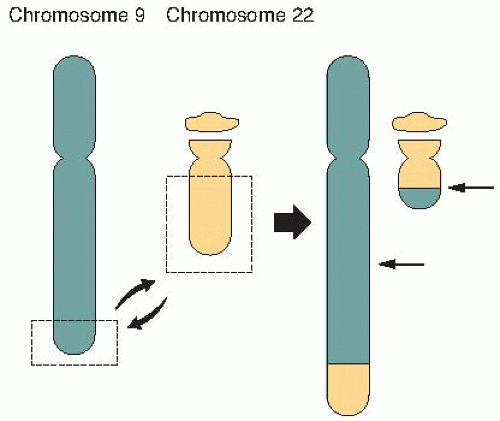
FIGURE 18.5 A symmetric translocation between chromosomes 9 and 22 brings together the bcl and abl genes to form a fusion gene associated with over 90% of cases of chronic myelogenous leukemia (CML).


TABLE 18.1 Examples of Chromosomal Changes Leading to Oncogene Activation and Their Associated Murine or Human Malignanciesa


Oncogene


Chromosomal Change


Cancer


int-1


Proviral insertion


Murine breast carcinoma


int-2


Proviral insertion


Murine breast carcinoma


pim-1


Proviral insertion


Murine T-cell lymphoma


N-ras


Point mutation (1)


Melanoma


K-ras


Point mutation (12)


Pancreas carcinoma


H-ras


Point mutation (11)


Colon carcinoma


neu


Point mutation (17)


Neuroblastoma


N-myc


Gene amplification (8)


Neuroblastoma


L-myc


Gene amplification (8)


Lung carcinoma


neu


Gene amplification (17)


Breast carcinoma


EGFR


Gene amplification (7)


Squamous cell carcinoma


bcr-abl


Translocation (9-22)


Chronic myelogenous leukemia


c-myc


Translocation (8-14)


Burkitt lymphoma


c-myc


Translocation (2-8)


Burkitt lymphoma


c-myc


Translocation (8-22)


Burkitt lymphoma


bcl-2


Translocation (14-18)


Diffuse large B-cell lymphoma


a Human oncogenes activated by retroviruses have not yet been found in human malignancies, only murine cancers.


▪ MUTATION AND INACTIVATION OF TUMOR SUPPRESSOR GENES


The Retinoblastoma Paradigm

Oncogenes result from a mutation, deletion, or alteration in the expression of one copy of a gene. Thus, oncogenes are dominant genes because a mutation in only one copy will cause their activation even though the other copy of the gene is unchanged. This concept led to speculation that another class of genes, termed “antioncogenes,” suppresses the effect of oncogenes on transformation and tumor formation. The existence of tumor suppressor genes was supported by cell fusion studies between tumor cells and normal cells and by the family history studies of people afflicted with inherited cancer-prone disorders such as retinoblastoma or Li- Fraumeni syndrome. Although one mutated version of an oncogene is sufficient to drive malignant progression, one functional copy of a tumor suppressor gene is sufficient to suppress transformation, suggesting that both copies of a tumor suppressor gene must be inactivated to inhibit tumor growth (Fig. 18.6). Insight into the mechanism of tumor suppressor gene inactivation came from Knudson’s epidemiologic studies of families in which retinoblastoma appeared to be inherited in an autosomal dominant manner. Patients with familial retinoblastoma develop bilateral or multifocal disease at an earlier age than
patients with sporadic retinoblastoma. Based on these observations, Knudson proposed that in the inherited form of retinoblastoma, patients possess a germ line mutation of the retinoblastoma (Rb) gene in all the cells of their body, but inactivation of one Rb allele does not give rise to retinoblastoma. Thus, the disease appeared to be autosomal dominant because individuals are born with one mutated allele. However, a second mutation in a retinal cell is required to develop retinoblastoma. In the sporadic form of the disease, an individual has to acquire two Rb mutations in the same retinal cell to develop retinoblastoma. The “two-hit hypothesis” by Knudson provided a genetic basis to understand the differences in inherited and sporadic mutations in the onset of tumors and to advance the concept that both alleles of a tumor suppressor gene need to be inactivated to promote tumor development. Thus, tumor suppressor genes are recessive genes that require the inactivation of both functional gene copies before malignancies develop, whereas loss of one functional copy results only in increased cancer susceptibility. In addition, it is often the case that the same tumor suppressor gene involved in hereditary cancer syndromes, such as retinoblastoma, is also inactivated in other forms of cancer. The Rb gene itself has now been implicated in several other human cancers, which indicates that it may play a generalized role in tumor growth suppression in various tissues. For example, patients who are cured of familial retinoblastoma are at increased risk of osteosarcoma, small cell lung cancer, and breast cancer; although the loss of the Rb gene alone is sufficient for retinoblastoma, further changes are required for the development of these other tumors.


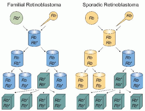
FIGURE 18.6 Rb mutations in familial and sporadic retinoblastoma. In familial retinoblastoma, one normal allele (Rb) and one mutated allele (Rb*) are inherited from either parent, resulting in a heterozygous individual containing Rb/Rb* retinal cells. Subsequent mutation in any retinal cell inactivates the remaining normal Rb allele, leading to loss of growth control and expansion of the homozygous mutant Rb*/Rb* retinal cells that leads to retinoblastoma. In sporadic retinoblastoma, two normal Rb alleles are inherited from each parent. First, a mutation inactivates one copy, resulting in heterozygous Rb/Rb* retinal cells. A subsequent mutation within the same retinal cell inactivates the remaining copy of normal Rb, leading to loss of growth control and expansion of homozygous Rb*/Rb* retinal cells that leads to retinoblastoma. (Illustrating the concepts proposed by Knudson AG. Mutation and cancer: statistical study of retinoblastoma. Proc Natl Acad Sci USA. 1971;68:820-823.)


The Li-Fraumeni Paradigm

The Li-Fraumeni syndrome (LFS) is a rare autosomal, dominantly inherited disease that predisposes individuals to develop osteosarcomas, soft-tissue sarcomas, rhabdomyosarcomas, leukemias, brain tumors, and carcinomas of the lung and breast (Fig. 18.7). Initial attempts to identify the genetic mutations that underlie LFS were unsuccessful because of the rarity of the syndrome and the high mortality of the patients. The major insight into the underlying cause of LFS came when it was found that mice that overexpress a mutant version of the p53 tumor suppressor gene in the presence of the wild-type p53 gene develop a spectrum of tumors similar to that seen in patients with LFS. Sequencing of p53 in affected family members revealed germ line missense mutations in the p53 tumor suppressor gene located on chromosome 17p13 that resulted in its inactivation, and tumors derived from affected individuals had lost the remaining wild-type allele of p53. Similar to retinoblastoma, loss or inactivation of both wild-type copies of p53 is needed for tumor formation. However, functional loss of one germ line inherited copy of mutant p53 accelerates the onset of spontaneous tumor formation. Therefore, patients with LFS follow a similar paradigm as patients with retinoblastoma in developing spontaneous tumors, but unlike retinoblastoma in which germ line mutations mainly give rise to retinal tumors, loss of p53 results in a wide spectrum of tumors. Table 18.2 provides examples of other cancer predisposition genes and their associated syndromes.


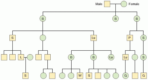
FIGURE 18.7 Pedigree analysis of familial cancer history of LFS. Symbols: B, breast cancer; G, glioblastoma; L, leukemia; Lu, lung cancer; P, pancreatic cancer; S, sarcoma; W, Wilms tumor. (Adapted from Li FP, Fraumeni JF. Prospective study of a family cancer syndrome. J Amer Med Assoc. 1982;247:2692-2694, with permission.)


Familial Breast Cancer, BRCA1 and BRCA2

The breast cancer susceptibility genes (BRCA1 and BRCA2) are found mutated in 5% and 10% of all breast cancer cases and are also associated with familial ovarian and prostate cancers. Familial breast cancer can be distinguished from sporadic breast cancer by its earlier age of onset, an increased frequency of bilateral tumors, and a higher incidence of cancer, in general, within an affected family. Histopathologically and anatomically, familial and sporadic cases of breast cancer are indistinguishable. Although BRCA1 and BRCA2 mutations account for the most inherited forms of breast cancer, they are rarely found in sporadic cases of breast cancer. BRCA1 and BRCA2 have been implicated in DNA damage, repair, cell cycle progression, transcription, ubiquitination, apoptosis, and in the determination of stem-cell fate. The most well-studied roles of BRCA1 and BRCA2 are in homologous recombination (see Chapter 2). Phenotypically, cells deficient in BRCA1 or BRCA2 exhibit elevated levels of genomic instability, which is consistent with their roles in homologous recombination. This conclusion is also supported
by the finding that mice lacking BRCA1 have the same phenotype as those null for the essential homologous recombination protein RAD51. Functionally, BRCA1 acts as a sensor of DNA damage and replication stress and mediates homologous recombination through BRCA2. In response to DNA damage-induced ionizing radiation, BRCA1 is phosphorylated at numerous sites by ataxia telangiectasia mutated (ATM), ataxia telangiectasia and Rad3-related (ATR), and checkpoint kinase 2 (Chk2) that direct it to associate with other repair proteins in nucleotide excision repair (NER), including xeroderma pigmentosum, complementation group C (XPC) and DNA damage-binding protein 2 (DDB2), in mismatch repair (MMR) MSH2, MSH6, and in DNA damage signalling ATM and the MRN complex (MRE11, Rad50, and NBS1). In contrast to BRCA1, BRCA2 appears to play a more direct role in homologous recombination by binding to RAD51 and aiding in the formation of RAD51 foci at the sites of DNA breaks. Cells from patients deficient in BRCA1 or BRCA2 are defective in homologous recombination and rely on error prone nonhomologous end- joining (NHEJ) to repair their DNA double strand breaks (DSBs). The resulting accumulation of mutations in BRCA1 and BRCA2 deficient cells promotes tumor formation.


TABLE 18.2 Examples of Cancer Predisposition Genes and Their Associated Syndromes


Tumor Suppressor Gene


Syndrome


Tumor


Rb


Retinoblastoma


Retinoblastoma


WT1


Familial Wilms tumor


Wilms tumor


NFI


Neurofibromatosis type 1


Neurofibroma, sarcoma


NF2


Neurofibromatosis type 2


Schwannoma, meningioma


APC


Familial adenomatosis polyposis


Tumor of colon, stomach, and intestine


p53


Li-Fraumeni syndrome


Breast, lung, brain tumors, sarcoma


VHL


von Hippel-Lindau disease


Tumor of kidney, adrenal


E-CAD


Familial gastric cancer


Tumor of stomach, breast


PTCH


Gorlin syndrome


Basal cell carcinoma


PTEN


Cowden syndrome


Hamartoma


MEN1


Multiple endocrine neoplasia


Tumor of pituitary, pancreas, and parathyroid


▪ SOMATIC HOMOZYGOSITY

How are recessive tumor suppressor genes lost? Cytogenetic studies are used to identify chromosomal changes in peripheral blood lymphocytes or fibroblasts from patients with cancer, especially those with a family history of cancer, to identify chromosomal rearrangements or deletions. At the subchromosomal level, a genomewide linkage analysis is used to determine that a certain chromosome region is tightly linked with cancer predisposition. Both copies of a suppressor gene in the sporadic form of retinoblastoma and other solid tumors may result from two independent allelic mutations, but in practice, it occurs more often by the process of somatic homozygosity. The steps appear to be as follows: One chromosome of a pair is lost, a deletion occurs in the remaining chromosome, and the chromosome with the deletion replicates. Instead of having each of the two alleles contributed by different parents, the cell has both alleles from the same parent, with loss of a vital piece containing the tumor suppressor gene (Fig. 18.8). This process has been documented for chromosome 13 in the case of retinoblastoma, chromosome 11 in Wilms tumor, chromosome 3 for small cell lung cancer, and chromosome 5 for colon cancer. Most interesting of all, perhaps, is the case of
astrocytomas in which somatic homozygosity is observed for chromosome 10 in grade II and III astrocytoma and for both chromosomes 10 and 17 for grade IV glioblastoma.


FIGURE 18.8 The process of somatic homozygosity. In a normal cell, there are two copies of each chromosome, one inherited from each parent. For a given suppressor gene to be inactivated, the copy must be lost from both chromosomes. This could, of course, occur by independent deletions from the two chromosomes; but in practice, it is more common for a single deletion to occur in one chromosome while the second chromosome is lost completely. The remaining chromosome, with the deletion, then replicates. The cell is thus homozygous, rather than heterozygous, for that chromosome.


▪ THE MULTISTEP NATURE OF CANCER

Perhaps the most pervasive dogma in cancer research is that carcinogenesis is a multistage process. The implication is that there are several distinct events that may be separated in time. This idea is almost 70 years old and is exemplified by the skin cancer experiments in mice that introduced the concepts of initiation, promotion, and progression as stages in tumor development.

Genetic analysis of cells from solid tumors also suggests alterations, mutations, or deletions in multiple signaling genes, either oncogenes or suppressor genes; 6 to 12 mutations have been suggested for the formation of a carcinoma. In the case of colorectal cancer, a model has been proposed that correlates a series of chromosomal and molecular events with the changes in the histopathology of normal epithelium during the multistage formation of colorectal cancer and metastatic carcinoma. This concept is illustrated in Figure 18.9.


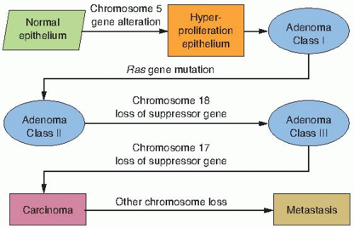
FIGURE 18.9 Cancer has long been thought to be a multistep process and has been described with operational terms such as initiation, promotion, and progression. In at least one human malignancy, namely, colon cancer, the molecular events during the progress of the disease have been identified. (Based on the work of Vogelstein and Kinzler.)

A more general model of the series of events in carcinogenesis is shown in Figure 18.10. The first event, from whatever cause (including ionizing radiation), causes a mutation in a gene in one of the families responsible for the stability of the genome. This leads to a mutator phenotype, so that with many cells dividing, multiple mutations are likely in cancer-associated genes, both oncogenes and tumor suppressor genes. This in turn leads to progression of the cancer and ultimately its invasive and metastatic properties.

Therefore, it is not surprising that restoration of one copy of a tumor suppressor gene is sometimes not sufficient to restore tumor suppressor activity because solid tumors accumulate mutations that can make them refractory to the restoration of a single tumor suppressor gene. Although tumor suppressor genes are functionally quite different from oncogenes, they modulate similar cellular targets as oncogenes.


FIGURE 18.10 Illustrating the multistep nature of carcinogenesis and the concept of the mutator phenotype. The first step in carcinogenesis by radiation or any other agent may be a mutation in one of the gene families responsible for the stability of the genome. This may be a DNA repair gene, an MMR gene, or a gene in a family as yet unidentified. This leads to the mutator phenotype, with multiple mutations possible in both oncogenes and tumor suppressor genes. This then leads to a series of steps that result in an invasive metastatic cancer. Not all the same mutations need to be present in every case.


▪ FUNCTION OF ONCOGENES AND TUMOR SUPPRESSOR GENES

The myriad of genetic and epigenetic changes that drive tumor evolution is a systems biology problem in which cells can be thought of as circuits, where an alteration of the circuit can lead to increased output, decreased output, complete loss of output, or no change. Therefore, cancer biologists attempt to determine how a specific gene, when mutated, alters normal tissue function. To understand how oncogenes and tumor suppressor genes lead to neoplasia, we need to understand how each of these circuits impacts normal cellular physiology. What cellular functions are disrupted by oncogene activation and tumor suppressor gene inactivation, and how do these disrupted functions affect the differentiation, growth, and death of cells? What follows is a description of the general categories of cell functions that are perturbed by deregulation of these two classes of genes during malignant progression.


Deregulated Proliferation

The loss of proliferative control of cancer cells is evident to all who study cancer. In fact, the earliest concept suggested by tumor biologists was that cancer was a disease of uncontrolled proliferation. Untransformed cells respond to extracellular growth signals known as mitogens through a transmembrane receptor that signals to intracellular circuits that increase growth. Thus, the growth factor, the receptor, and the intracellular circuits can all lead to self-sufficiency when deregulated. Typically, one cell secretes a mitogenic signal to stimulate the proliferation of another cell type. For example, an epithelial cell can secrete a signal to stimulate fibroblasts to proliferate. In contrast to untransformed cells, transformed cells have become autonomous in regulating their growth by responding to the mitogenic signals they themselves produce. In this manner, they use an autocrine circuit to escape the need for other cell types. For example, mesenchymal cells are responsive to transformation by v-sis, because they possess receptors for platelet derived growth factor (PDGF), and breast epithelial cells are responsive to int-2, because they possess receptors for fibroblast growth factor (FGF).

If overexpression of growth factors can lead to uncontrolled proliferation, then continuous activation or overexpression of growth factor receptors will do the same. Several well-known oncogenes, such as v-erb-2 (HER-2/neu) and v-fms, encode growth factor receptors. These receptors are mutated at their amino terminal
residues so that they no longer require their respective growth factor (ligand) to signal induction of their kinase activity. Growth factor receptors can structurally be divided into extracellular ligand-binding domains (LBDs), transmembrane- spanning domains (TMS), and intracellular kinase domains. Although mutations have been found in all three domains, mutations in the ligand-binding domains are a common alteration that results in constitutive kinase activity that transduces the signal for the cell to proliferate. In addition to structural alterations in the receptor, some tumors overexpress growth factor receptors that make them hyperresponsive to physiologic levels of growth factor stimulation. In contrast to mitogenic-responsive growth factor receptors, a second class of receptors that transmit signals from the extracellular matrix can also regulate proliferation. Integrin receptors are the prototypical example of this class of regulators that transmit signals from different components of the extracellular matrix (ECM) to signal proliferation or quiescence.

There are numerous intracellular circuits that transduce the signal from the cell surface to the nucleus of the cell. The src, ras, and abl proteins are all members of this group. Most members of this group are tyrosine kinases or serine/threonine kinases. Src

Only gold members can continue reading. Log In or Register to continue

Stay updated, free articles. Join our Telegram channel

Jun 1, 2016 | Posted by in GENERAL RADIOLOGY | Comments Off on Cancer Biology

Full access? Get Clinical Tree

Get Clinical Tree app for offline access