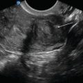
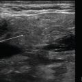
Figure 3.1
Parasternal long transducer placement: Place the transducer on the anterior chest at the second or third intercostal space to the left of the sternum with the transducer marker toward the patient’s right shoulder
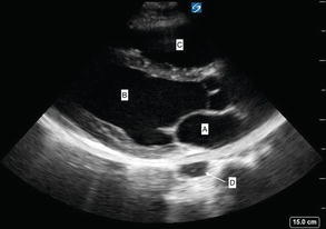
Figure 3.2
Parasternal long: Parasternal long cardiac view in between cardiac cycles with both mitral and aortic valves closed. Blood flows from the left atrium (a) through the mitral valve, into the left ventricle (b), and out to the body through the aortic valve. The right ventricle can be seen at the top of the screen (c). The descending thoracic aorta is posterior to the heart (d)
Parasternal Short Axis
Transducer Placement.
From the long axis position, rotate the transducer approximately 90° clockwise so the marker points toward the left shoulder.
This will give you a cross-sectional view of the left ventricle.
Figure 3.3—Parasternal short transducer placement.
Ultrasound Anatomy.
Figure 3.4—Parasternal short
Left ventricle appears as a muscular circle that, in the absence of pathology, contracts concentrically.
The bicuspid mitral valve is located centrally within the left ventricle that has the appearance of a “fish mouth” opening and closing.
Figure 3.5—Mitral valve fish mouth in parasternal short
Video 3.2—Parasternal short at the mitral valve
Sliding the probe toward the apex will allow visualization of the two papillary muscles in short axis.
Each papillary muscle is usually located at 4 and 8 o’clock within the circular left ventricle.
Video 3.3—Parasternal short at the apex.
Right ventricle will appear as a “C”-shaped structure on the left side of the screen, separated from the left ventricle by the septal wall.
Myocardial infarction or ischemia can be identified by wall motion abnormalities and a loss of concentric contraction of the left ventricle.
Anterior wall is closest to the transducer.
Septal wall is to the left of the screen.
Lateral wall is to the right of the screen.
Interior wall is in the far field.
Figure 3.6—Walls of LV labeled in parasternal short.

Figure 3.3
Parasternal short transducer placement: From the long axis position, rotate the transducer approximately 90° clockwise so the marker points toward the left shoulder

Figure 3.4
Parasternal short labeled: Parasternal short cardiac view at the level of the papillary muscles (arrows). LV cavity (a) is circular, not elongated or ovoid. The RV cavity (b) can be seen in upper left

Figure 3.5
Mitral valve fish mouth in parasternal short: Mitral valve parasternal short view displaying the “fish mouth” of the mitral valve in short axis. The anterior leaflet (a) can be seen above the posterior leaflet (b)

Figure 3.6
Walls of LV labeled in parasternal short: Parasternal short cardiac view at the level of the papillary muscles showing the relative wall sections
Apical Four- and Two-Chamber
- (a)
Transducer Placement
Place the transducer on the point of maximal impulse (PMI) with the marker pointed toward the patient’s left elbow.
Figure 3.7—Apical four-chamber transducer placement.
This will allow visualization of the four chambers of the heart, viewed from apex to base.
Rotate the transducer counterclockwise to obtain a two-chamber view of the left atrium and left ventricle.
- (b)
Ultrasound Anatomy
Four-chamber
Figure 3.8—Apical four-chamber ultrasound image.
Left ventricle and atrium will be seen on the right side of the screen.
The left ventricle wall on the far right is the lateral wall.
The septal wall separates the two ventricles and is in the middle of the screen in a vertical orientation.
Right ventricle and atrium will be seen on the left side of the screen.
Video 3.4—Apical four-chamber.
Two-chamber
Figure 3.9—Apical two-chamber.
Only the left ventricle and left atrium will be seen in this view.
This view allows visualization of the anterior and inferior walls of the left ventricle.
Video 3.5—Apical two-chamber.

Figure 3.7
Apical four-chamber transducer placement: Place the transducer on the point of maximal impulse (PMI) with the marker pointed toward the patient’s left elbow

Figure 3.8
Apical four-chamber: Apical four-chamber view in systole, with apex of the heart located at the top of the screen, base located at the bottom. Ideal orientation is with septal wall (between d and b) located in vertical orientation, perpendicular to the face of the cardiac transducer. Cardiac chambers are as follows: left atrium (a), left ventricle (b), right atrium (c), and right ventricle (d)

Figure 3.9
Apical two-chamber: Apical two-chamber cardiac view demonstrating vertical orientation of the heart. Inferior wall of the left ventricle (b) is to the left of the image, anterior wall to the right. Left atrium (a) and aortic outflow (c) are also demonstrated
Subxiphoid
Transducer Placement
Place the transducer just below the xiphoid process with the marker pointed toward the patient’s left.
Place operator’s hand on top of the transducer, use a moderate amount of force to press the transducer into the patient’s abdomen, and angle the transducer superior and leftward toward the left shoulder.
Adjust depth to adequately visualize the entire heart.
Figure 3.10—Subxiphoid transducer placement.
Ultrasound Anatomy
Figure 3.11—Subxiphoid ultrasound image.
The liver is anterior to the heart.
Posterior to the liver is the right ventricle and right atrium.
Posterior to the right heart is the left ventricle and left atrium.
Video 3.6—Subxiphoid.

Figure 3.10
Subxiphoid transducer placement: Place the transducer just below the xiphoid process with the marker pointed toward the patient’s left

Figure 3.11
Subxiphoid: Subxiphoid cardiac view demonstrating the heart viewed through the liver window. Cardiac chambers are identified as follows: left atrium (a), left ventricle (b), right atrium (c), right ventricle (d)
Inferior Vena Cava
- (a)
Transducer Placement
Place the transducer just below the xiphoid process, slightly to the right of midline, with the marker pointed toward the patient’s head.
The transducer should be perpendicular to the spine.
Figure 3.12—IVC transducer placement.
- (b)
Ultrasound AnatomyGet Clinical Tree app for offline access
Figure 3.13—IVC ultrasound image.
IVC will be seen posterior to the liver entering the right atrium.
Video 3.7—IVC
IVC normally collapses with inspiration and dilates with expiration.
Dilated IVC with minimal collapse indicates volume overload; impaired cardiac filling, such as with tamponade; outflow obstruction, such as with PE; or cardiogenic shock.
Figure 3.14—Dilated IVC
Video 3.8—Dilated IVC
Increased IVC collapse may indicate hypovolemia.
Figure 3.15—Collapsed IVC
Video 3.9—Collapsed IVC

Stay updated, free articles. Join our Telegram channel

Full access? Get Clinical Tree


