, J. Stoitsis2 and K. S. Nikita2
(1)
School of Medicine, University of Athens, Athens, Greece
(2)
Electrical and Computer Engineering, Technical University of Athens, Athens, Greece
Abstract
It has been shown that computerized analysis of ultrasound images of the carotid artery may provide quantitative disease biomarkers and can potentially serve as a ”second opinion” in the diagnosis of carotid atherosclerosis. Extending the findings of previous work on the subject, a set of methodologies are presented in this chapter, suitable for application on two-dimensional B-mode ultrasound images. More specifically, a Hough-Transform-based technique for automatic segmentation of the arterial wall allows the estimation of the intima-media thickness and the arterial distension waveform, two widely used determinants of arterial disease. Texture features extracted from Fourier-, wavelet-, and Gabor-filter-based methods can characterize symptomatic and asymptomatic atheromatous plaque. Finally, a methodology based on least-squares optical flow is proposed for the analysis and quantification of motion of the arterial wall.The suggested methodologies allow the extraction of useful biomarkers for the study of (a) the physiology of the arterial wall and (b) the mechanisms of carotid atherosclerosis.
1 Introduction
The carotid arteries are responsible for supplying blood to the brain. Each common carotid artery divides into an external and an internal branch at the carotid bifurcation. The external carotids supply blood to the neck, pharynx, larynx, lower jaw and face, whereas the internal carotids enter the skull delivering blood to the brain. The presence of an atheromatous lesion, or plaque, in the carotid arteries, also known as carotid atherosclerosis, may disturb the normal circulatory supply to the brain. Carotid atherosclerosis may produce total occlusion of a specific arterial site or cause a thromboembolic event. In advanced stages of the disease, cerebrovascular symptoms, such as transient ischaemic attack, amaurosis fugax (temporal blinding) or stroke, may occur.
Ultrasound imaging of the carotid artery is the most widely used modality in the diagnosis of carotid atherosclerosis due to its noninvasiveness, non-ionizing nature and low cost. In particular, B (Brightness)-mode imaging, i.e. the reproduction of the amplitude of the reflected waves by their brightness, is commonly used to assess arterial wall morphology. B-mode images exhibit a granular appearance, called speckle pattern, which is caused by the constructive and destructive interference of the wavelets scattered by the tissue components. In B-mode ultrasound, blood reflects very little and the vessel lumen appears as a hypoechoic band. Figure 1 shows examples of B-mode ultrasound images of (a) a healthy (non-atherosclerotic) and (b) a diseased (atherosclerotic) carotid artery.
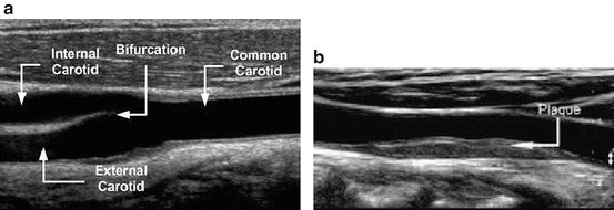

Fig. 1
Examples of B-mode ultrasound images of (a) a healthy (non-atherosclerotic) and (b) a diseased (atherosclerotic) carotid artery
2 Quantitative assessment of carotid atherosclerosis
Currently, severity of carotid atherosclerosis and selection of patients to be considered for endarterectomy, i.e. surgical removal of plaque, are based (a) on the degree of stenosis caused by the plaque, in asymptomatic subjects, and (b) on both the degree of stenosis and previous occurrence of clinical symptoms, in symptomatic subjects. However, there is evidence that atheromatous plaques with relatively low stenosis degree may produce symptoms and that highly stenotic atherosclerotic plaques can remain asymptomatic. Because not all carotid plaques are necessarily harmful and because carotid endarterectomy carries a considerable risk for the patient, the crucial task of optimized selection of patients for operation may be greatly facilitated by the use of novel biomarkers. B-mode ultrasonic images, in combination with appropriate image processing methods for segmentation, texture and motion analysis, may be used to extract useful diagnostic indices of the geometry, echogenicity and strain, respectively, of the carotid artery wall.
2.1 Prior Art
Previous work on computerized analysis of ultrasound images of the carotid arteries includes automatic segmentation of the arterial lumen, plaque texture analysis and tissue motion analysis. The use of deformable models [12], including snakes [3], allows automatic identification of the random-shaped carotid artery wall from static ultrasound images. In addition to this, the Hough Transform (HT) has been used to segment the arterial wall from sequences of images [15, 17]. In this case, the arterial distension waveform can be estimated facilitating the study of the dynamic arterial geometry. Plaque echogenicity, estimated from B-mode ultrasound images using texture analysis techniques, may be used to characterize atheromatous plaque and differentiate between symptomatic and asymptomatic cases. Plaque echogenicity has been analyzed using a number of statistical, model-based and Fourier-based methods [4, 16]. Motion of the arterial wall and atheromatous plaque has recently gained attention as a determinant of carotid atherosclerosis. It has been shown that plaque strain, expressed as relative motion between different parts of the plaque, may be related to plaque instability, i.e. to the risk for cerebrovascular complications, such as stroke [13]. Temporal sequences of ultrasound images can be used to estimate movement of the carotid artery wall by tracking the speckle patterns generated by the tissue [5, 7, 13].
The purpose of this chapter is to suggest a set of methodologies which extend the findings of previous work on computerized analysis of ultrasound images of the carotid artery in an attempt to identify quantitative indices of diagnostic value. Such indices may be useful for early and valid diagnosis of atherosclerosis as well as for the study of the physiology of the normal and diseased arterial wall.
3 Computerized analysis of ultrasound images of carotid artery
The methodologies described in this chapter are suitable for two-dimensional (2D) B-mode ultrasound imaging. This is the most widely used modality for the assessment of the carotid artery. The methodologies aim at (a) automatic segmentation of the arterial wall from longitudinal and transverse sections, (b) texture analysis of atheromatous plaque and (c) analysis and quantification of motion of the arterial wall. It is recommended that the methodologies be applied to normalized ultrasound images, according to widely accepted specifications [6], to minimize variability introduced by different equipment, operators and gain settings and facilitate imaged tissue comparability. The techniques are designed to be applied to sequences of images, thus allowing the extraction of quantitative information at different phases of the cardiac cycle, e.g. systole or diastole. Obviously, it is possible to apply them to individual (static) images, if this is required by the clinical application.
4 Early disease biomarkers
Early disease stages may be assessed by interrogating the arterial wall on which focal lesions (plaques) may not yet have become obvious. The suggested HT technique allows the automatic extraction of straight lines and circles to approximate the wall-lumen boundary in longitudinal and transverse sections, respectively. HT can be used to detect parametric curves of the form  in digital images, where c is the vector of coordinates, p the vector of parameters and i = 1. . . n, the number of parameters required to define the curve. HT transforms the image to an n-dimensional parametric space, called the accumulator array. Operating on a binary image of edge pixels, all possible curves v(c, p i ) = 0 through a pixel with vector coordinates c are transformed to a combination of parameters p i , which then increment the corresponding cell of the accumulator array. The main steps of the methodology, which are described in detail in [8], include:
 in digital images, where c is the vector of coordinates, p the vector of parameters and i = 1. . . n, the number of parameters required to define the curve. HT transforms the image to an n-dimensional parametric space, called the accumulator array. Operating on a binary image of edge pixels, all possible curves v(c, p i ) = 0 through a pixel with vector coordinates c are transformed to a combination of parameters p i , which then increment the corresponding cell of the accumulator array. The main steps of the methodology, which are described in detail in [8], include:
Reduction of image area
This may be achieved by automatically isolating a rectangular area containing the vessel lumen. To this end, four points may be defined to delimit the area to be investigated. This is an important step because it minimizes the possibility to detect unwanted structures, which may be present biasing the representation of the arterial lumen and, thus, reduces the computational cost and the time required to perform the segmentation task.
Image pre-processing
This step includes removal of high frequency noise using a symmetric Gaussian lowpass filter and morphological closing to suppress small ‘channels’ and ‘openings’ of the image.
Edge detection
The image is first transformed into binary through the application of a global threshold and then a Sobel gradient operator may be applied.
Hough Transform
Longitudinal sections are searched for lines defined as  , where z is the distance from the left upper corner of the image and θ is the angle with the x-axis. Transverse sections are searched for circles defined as
  , where (a, b) are the coordinates of the center and r is the radius of the circle.
 , where z is the distance from the left upper corner of the image and θ is the angle with the x-axis. Transverse sections are searched for circles defined as
  , where (a, b) are the coordinates of the center and r is the radius of the circle.
Selection of dominant lines/circle


Two lines in longitudinal sections and one circle in transverse sections with the maximal values in the corresponding accumulator arrays are eventually selected, representing the boundaries of the wall-lumen interface.
Stay updated, free articles. Join our Telegram channel

Full access? Get Clinical Tree


