5 Carpal Tunnel Evaluation and Injection
♦ Setup
• The patient is seated, facing the operator, with the dorsal forearm resting on the table.
• The elbow is flexed to about 45 degrees.
• A probe with a high frequency is used. High-frequency probes give greater detail but do not penetrate deeply.
♦ Landmarks
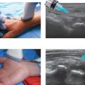
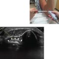
The landmarks are identified visually and with palpation (Fig. 5.1):
• Distal wrist crease
• Pisiform

Fig. 5.1 The pisiform is identified with palpation, and the distal wrist crease is visualized.
♦ Probe Positioning
Axial
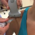
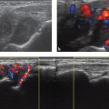
• The probe is centered over the distal wrist crease (Fig. 5.2). It should be perpendicular to the forearm.
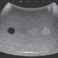
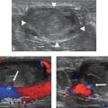
• This view is useful for injections and measurement of the cross-sectional area of the median nerve. It can also be used for evaluation of ganglion cysts or masses within the carpal tunnel (Videos 5.1 and 5.2).

Fig. 5.2 (a) The probe is positioned 90 degrees to the long axis of the forearm. (b) Ultrasound image of the carpal tunnel. The median nerve is circled in red.
Related posts:
Stay updated, free articles. Join our Telegram channel

Full access? Get Clinical Tree


