Susie J. Park • Gary P. Siskin
Throughout this textbook, there are several chapters that focus on the embolic agents used for embolization and many more chapters focusing on the clinical indications being treated with embolization procedures. There is no doubt that these two broad areas of focus are essential for these procedures to be successfully performed. However, there is a third area of focus that requires attention as well: the catheters and the catheterization techniques used today for embolization. Many times, embolization procedures are either successful or unsuccessful based on the choices made by individual physicians regarding the materials and techniques used to deliver an agent to its intended target. This chapter will take a unique look at embolization from this perspective.
CATHETERS USED FOR EMBOLIZATION
Almost every available angiographic catheter can be used to deliver one or more of the many agents available today for embolization. Standard 4-Fr and 5-Fr angiographic catheters can be used to deliver coils, plugs, and particulate embolic agents. However, the expanded indications for embolization into smaller vessels that are increasingly located distally within a target vascular bed are requiring the use of microcatheters. Microcatheter characteristics that are favorable when it comes to the performance of embolization procedures include trackability, flexibility, kink resistance, and radiopacity; radiopacity is important to ensure that an embolic agent will be deployed exactly where deployment is intended to be. Many microcatheters are being manufactured with various tip configurations to facilitate catheterization of target vessels. In addition, microcatheters are now available with various inner and outer diameter sizes, so care must be taken to ensure that the microcatheter selected is compatible with the agent being planned for embolization. Selective catheterization in the setting of embolization can be challenging at times, and almost nothing can be more frustrating for an interventionalist than to find out that the microcatheter used cannot accommodate a particular coil or microsphere after it has been positioned appropriately within the target vasculature.
CATHETER AND EMBOLIC AGENT COMPATIBILITY
Every embolic agent reviewed in the previous chapters requires a catheter for delivery to the vascular bed being targeted for embolization. The actual catheter selected for delivery will, of course, depend on the embolic agent being used and the degree of selective catheterization required for the condition being treated. It will also depend on the inner diameter of the selected catheter because different agents either require or are more easily delivered through appropriately sized catheters. There are some specific issues surrounding catheter selection and the agent being used for embolization.
Coils
When using coils for embolization, the delivery catheter must have an inner diameter that constrains the coil as it is being delivered. If the inner diameter of the catheter is too large relative to the size of the coil, then the potential exists for the coil to begin forming within the catheter. This can result in the coils getting stuck within the catheter, forcing the catheter to be removed. If catheterization of the target vessel was technically challenging, then being forced to remove the delivery catheter could have significant implications in terms of the additional radiation dose, contrast volume, and physician time required to repeat that portion of the procedure. This is particularly the case with the use of microcatheters and newer detachable coils. For example, the hydrogel coating of an AZUR detachable coil (AZUR Peripheral HydroCoil Embolization System; Terumo Medical Corporation, Somerset, New Jersey) may make it better placed with a microcatheter with a 0.027-in inner diameter as compared with other coils that are easily placed through a microcatheter with a 0.021-in inner diameter. Therefore, attention must be paid to catheter selection to avoid this type of catheter–coil mismatch.
Coil passage through a catheter can be held up for other reasons as well. It can occur with retrograde blood flow into the catheter with subsequent thrombosis. This can be prevented with frequent flushing of the catheter or even the use of a continuous infusion of saline through the catheter via a Tuohy Borst adapter (Cook Incorporated, Bloomington, Indiana).1 In addition, hydrophilic catheters can be softer than other catheters, which may cause fibered coils to get stuck in the catheter near a curve or the tip.2 This can result in a failure to deliver the coil to its target and the need to prematurely remove the obstructed catheter.
Catheter stability is an important component of coil delivery to prevent incorrect positioning of the coil as it is being deployed. The use of a guide catheter or long sheath can assist with minimizing this potential technical complication. In addition, before the use of any pushable coil, it is good practice to test the stability of the delivery catheter by either injecting saline through the catheter or by advancing the coil pusher to the tip of the catheter before any coil is placed.3
Plugs
It is important to understand that the available plugs typically require larger catheters for delivery when compared with coils, and they cannot be delivered with a microcatheter. For example, depending on the exact configuration and size of an Amplatzer Vascular Plug (St. Jude Medical, Inc., St. Paul, Minnesota), either a 4-Fr to 5-Fr catheter or 4-Fr to 7-Fr guide catheter/sheath would be required for delivery.4 Therefore, this understandably requires that the target vessel can accommodate a catheter or sheath of this size. It may or may not be possible for these catheters to track over a guidewire to an area of arterial pathology if the vessels leading to the pathology are small, tortuous, or diseased. This must be recognized before settling on the use of a plug for embolization.
Liquid Agents
Cyanoacrylates are one example of a liquid adhesive used for embolization procedures, and the main pathology treated with these agents are arteriovenous malformations. Glue embolization is typically performed through a microcatheter positioned as close as possible to the nidus.5 Because polycarbonate can be destroyed by cyanoacrylate, polypropylene syringes should be used for these procedures.5,6 Although attention to the technical details of administration is important to prevent catheter adhesion to intraluminal glue (e.g., dilution, catheter position, etc.), there is no specific microcatheter that is recommended for use with cyanoacrylates.
This is not the case with Onyx (Covidien, Irvine, California), which is another liquid embolic agent. Onyx is an ethylene vinyl alcohol copolymer used in conjunction with dimethyl sulfoxide (DMSO). Onyx is nonadhesive, which means that the delivery catheter is unlikely to become adhered to it after delivery. DMSO is necessary during Onyx administration because it inhibits the polymerization of Onyx, which begins after the diffusion of DMSO in the presence of aqueous media such as blood.7 The problem is that DMSO can also potentially erode a delivery catheter. As a result, the microcatheters used in association with DMSO administration must be DMSO compatible, indicating that the catheter manufacturer has done the appropriate testing to ensure that the catheter will maintain its integrity during DMSO and Onyx administration. The manufacturer of Onyx has done their own internal testing leading to specific recommendations for which catheters should be used with DMSO. The testing they perform is extensive, including detachment force testing (to determine the amount of force required to detach a catheter from Onyx), dynamic pressure testing (to determine the required pressure to push Onyx through a patent catheter), precipitated Onyx testing (to determine the pressure required to dislodge precipitated Onyx from both sides of the catheter), fuse joint segment burst testing (to determine the pressure required to burst each catheter-fused joint segment and the distal segment with marker band[s]), and others including catheter tensile strength testing, static burst testing, pressure profile testing, and collapsed lumen (kink) testing (Covidien, Laci Costa, written personal communication). As a result of this testing, recommended catheters include the Apollo, Marathon, UltraFlow, Echelon, and Rebar catheters, all of which are manufactured by Covidien (Irvine, California). There are other manufacturers claiming DMSO compatibility with their microcatheters as well, but the manufacturer of Onyx has not tested these catheters.
Particles
Particulate polyvinyl alcohol (PVA) particles have historically been associated with microcatheter clogging. This is most likely due to particulate aggregation, making the effective size of the PVA particles larger than their actual size.8 Selecting a catheter with an appropriate inner diameter is essential to ensure that catheters do not become occluded while using PVA particles. Although 4-Fr and 5-Fr catheters can certainly be used to administer PVA particles, it is more common practice for microcatheters to be used for this purpose. This is due to the vascular beds or tumors and other pathology that are typically targeted with PVA particles. In general, microcatheters with inner diameters of 0.027 to 0.028 in are more appropriate and easier to use in association with particulate embolization than smaller inner diameter catheters. Although it is possible to use microcatheters with smaller inner diameters, more injection force and more frequent flushing are required to avoid catheter occlusion. From a delivery perspective, the development of spherical embolic agents in recent years, including bland and drug-eluting microspheres, has resulted in easier administration and a lower incidence of microcatheter occlusion due to the fact that these microspheres do not aggregate.
Gelfoam
Gelatin sponge particles are commonly used for embolization. Interventionalists often prepare these particles at the time of the embolization procedure with the use of a cutting method or pumping method.9 Although gelfoam is most easily administered through a 4-Fr to 5-Fr angiographic catheter, it can be administered through a microcatheter as well. Administering gelfoam through a microcatheter can be done with the use of smaller syringes and higher injection pressures. However, concern has been raised that this can result in fragmentation of the gelfoam, leading to smaller particles being administered. Katsumori and Kashara10 as well as Osuga et al.11 have shown that this is not the case and that gelfoam can be safely delivered intact through a microcatheter.
CONSIDERATIONS FOR CATHETERIZATION DURING EMBOLOTHERAPY
Vascular Anatomy
During all embolization techniques, a thorough knowledge of vascular anatomy is required before embolization. Understanding the tissues and organs being supplied by individual vessels allows the interventionalist to increase the level of certainty that the target organ is being embolized and to gauge the risk of nontarget embolization. In addition, it is well established that there are various branching patterns of vessel origins all falling under the heading of “normal anatomy.” Without understanding where anomalous vessels to particular organs may arise, it would be impossible to be certain that all of the vessels responsible for the arterial supply are accounted for and embolized as necessary. For example, it has been well established that branches of the internal mammary artery can supply lung parenchyma in a patient with hemoptysis, that branches of the ovarian artery can supply the uterus in a patient with uterine fibroids, and that branches of the superior mesenteric artery can supply the liver in a patient with hepatocellular carcinoma. Having this knowledge can enable the interventionalist to study all possible sources of arterial supply to a target organ before concluding that the procedure has been successfully completed.
Nonselective versus Selective Catheterization
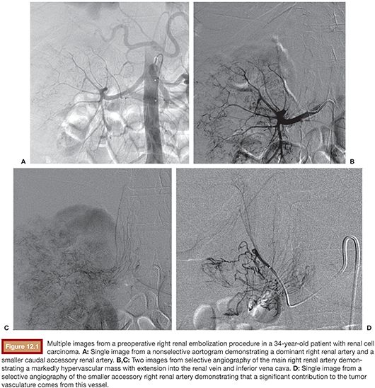
As part of the angiographic assessment before embolization, a nonselective angiogram is often performed to gain an overview of the arterial supply to the specific part of the body being targeted for embolization. There are several potential benefits to performing nonselective angiography before selective catheterization and embolization. Nonselective angiography permits an assessment regarding which vessels are actually responsible for supplying a target organ, which can eliminate or greatly reduce the time spent catheterizing vessels that are ultimately found to not be involved in the pathology being treated. In addition, the variant anatomy described in the previous section can be quickly identified so as not to allow for time being spent on the catheterization of noncontributory vessels. Nonselective angiography also allows for an assessment of those vessels that do require selective catheterization and can provide information to simplify the process of selective catheterization. Abdominal aortography, for example, can identify the number of renal arteries before selective renal embolization and which vessels are contributing to the abnormal target vasculature (Fig. 12.1). It can also identify the angle at which a renal artery originates from the abdominal aorta and can allow for appropriate selection of a catheter configuration that closely mimics the patient’s anatomy. This can reduce the time needed for selective catheterization, which in turn can limit the radiation dose and volume of contrast administered during this portion of an embolization procedure.
Stay updated, free articles. Join our Telegram channel

Full access? Get Clinical Tree


