Cecum is distended and lumen narrowed by medial folding and displacement, without a twist
IMAGING
• Radiography: Dilated, air-filled cecum in LUQ or abdominal midline
Single, long air-fluid level within cecum (upright or decubitus film)
Moderately distended gas or fluid-filled small bowel, little gas in distal colon
Markedly dilated cecum that appears upside down and backward with ileocecal valve directed laterally
• Additional CT signs
Whirl sign: Tightly twisted colonic wall and ileocolic mesenteric vessels
• Best imaging test: CT in axial and coronal planes
TOP DIFFERENTIAL DIAGNOSES
• Sigmoid volvulus
• Acute ileus
• Distal colon obstruction
• Ogilvie syndrome
PATHOLOGY
• Embryology/anatomy
Right colon is incompletely fused to posterior parietal peritoneum
• There are many causes of colonic distention, but ligamentous laxity is necessary for cecal volvulus to occur
CLINICAL ISSUES
• Accounts for 1/3 of colonic volvulus cases
• Complications: Ischemia, necrosis, perforation
• Treatment: Colonoscopy to reduce volvulus, surgery to prevent recurrence
DIAGNOSTIC CHECKLIST
• Rule out ileus, Ogilvie syndrome
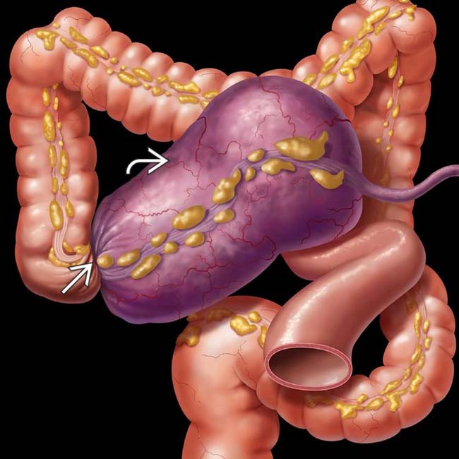
(Left) Graphic shows a twist (volvulus) of the ascending colon, obstructing the lumen and blood supply. The cecum on the mesentery is dilated and displaced toward the left upper quadrant (LUQ).
(Right) Supine radiograph shows a gas-distended segment of bowel (cecum) within the mid abdomen. The base of the cecum is directed toward the upper quadrant, and the ileocecal valve is directed laterally. Small bowel is gas-distended, whereas the left colon is relatively collapsed.
(Left) Axial CT in the same case shows a markedly distended cecum and a twisted (“whirled”) ileocolic mesentery within the right lower quadrant.
(Right) Coronal reformatted CT in the same case shows the markedly distended midline cecum . The cecum is upside down and backward, with the ileocecal valve pointed laterally. Coronal-reformatted CT is usually the most definitive study to show the characteristic features of cecal volvulus.
TERMINOLOGY
Synonyms
• Volvulus of cecum and part of ascending colon
Definitions
• Rotational twist of right colon on its axis, resulting in progressive distention and potential ischemia
• Cecal bascule
Cecum is distended and lumen narrowed by medial folding and displacement, without twist
IMAGING
General Features
• Best diagnostic clue
Markedly dilated cecum that appears upside down and backward with ileocecal valve directed laterally
Radiographic Findings
• Radiography
Dilated, air-filled cecum in left upper quadrant (LUQ) or abdominal midline
Only gold members can continue reading. Log In or Register to continue


 of the ascending colon, obstructing the lumen and blood supply. The cecum
  on the mesentery is dilated and displaced toward the left upper quadrant (LUQ).
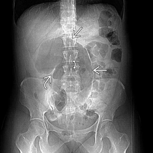
 within the mid abdomen. The base of the cecum is directed toward the upper quadrant, and the ileocecal valve
  is directed laterally. Small bowel is gas-distended, whereas the left colon is relatively collapsed.
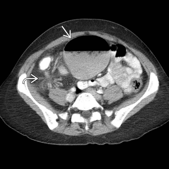
 and a twisted (“whirled”) ileocolic mesentery
  within the right lower quadrant.
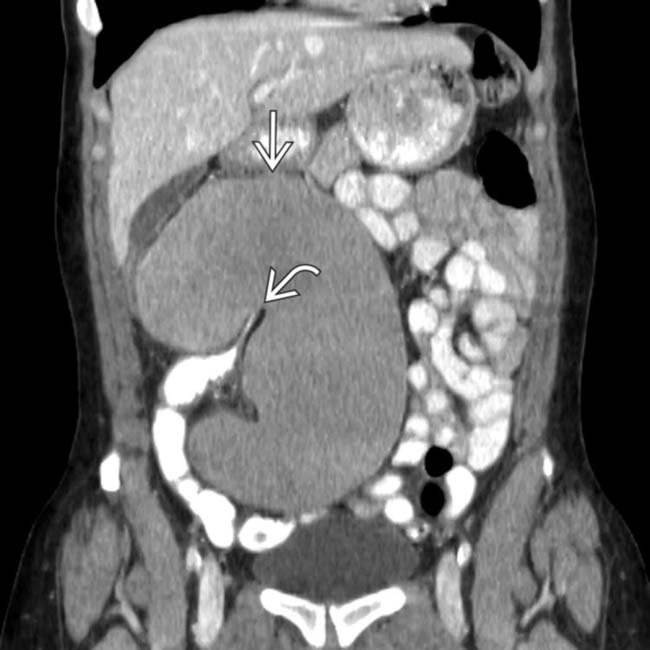
 . The cecum is upside down and backward, with the ileocecal valve
  pointed laterally. Coronal-reformatted CT is usually the most definitive study to show the characteristic features of cecal volvulus.


