CHAPTER 16 Cerebellum
The cerebellum (little brain) is the dorsal portion of the metencephalon situated within the posterior fossa. It is composed of three lobes: the anterior lobe, posterior lobe, and flocculonodular lobe.
ANATOMY
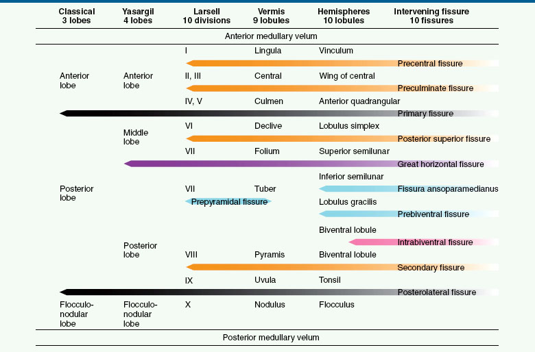
Grossly, the cerebellum is a small structure situated posterior and inferior to the bulk of the cerebrum (Fig. 16-1). It constitutes approximately 10% of the brain by weight but contains about half as many neurons as the cerebral hemispheres. The three lobes of the cerebellum contain nine lobules, distributed with three lobules in the anterior lobe, five in the posterior lobe, and one in the flocculonodular lobe. In most cases, the lobules of the vermis merge laterally with the lobules of the hemispheres. Along the posterior surface of the cerebellum, however, there is a slight offset in the alignment of the medial vermal lobules with the corresponding lateral hemispheric lobules (Fig. 16-2). The lobes and lobules have been classified in many ways. For the purposes of comparative anatomy, the Larsell classification is most specific.1 For clear medical communication, the common names are more useful. These are both provided, with synonyms, in Table 16-1.
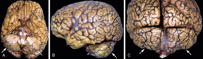
FIGURE 16-1 Relationships of the cerebrum to the cerebellum. Formalin-fixed human brain. A, Base view. B, Lateral view. C, Posterior view. The dural folds of the falx and tentorium have been removed. The cerebellum (little brain) (white arrows) lies posteroinferior to the cerebrum in relation to the inferior surfaces of the posterior temporal and occipital lobes. The thin “parallel” folia and fissures of the cerebellum are distinctly different from the swirling gyri and sulci of the cerebrum. The large vessels of the cerebellar surface course nearly perpendicular to the lines of folia and fissures. In C, the glistening arachnoid marks the posterior wall of cisterna magna. The posterior cerebellar notch of the cerebellum often does not align perfectly with the interhemispheric fissure above.
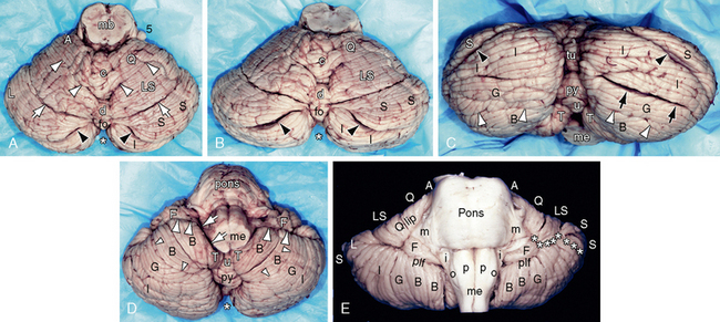
FIGURE 16-2 The surfaces, lobes, and lobules of the cerebellum. Two specimens. Removal of the pia-arachnoid and vessels exposes the pattern of the folia, fissures, and lobules. The major fissures have been opened by blunt finger dissection to emphasize their position and configuration. A to D, Specimen 1. A and B, The superior (tentorial) surface seen from directly above (A) and from above and behind (B). The superior surface has an overall butterfly shape, with a midline vermis situated between flanking cerebellar hemispheres. The back of the midbrain (mb) is enclosed by the anterior cerebellum. The anterior angles (A) mark the anteriormost extent of the cerebellum and lie just above and behind the sites at which CN V (trigeminal nerve) (5) enters the pons. The lateral angles (L) mark the lateral extremes of the hemispheres. The anterolateral and posterolateral margins separate the superior surface from the anterior and posterior surfaces. The posterior surface of the vermis is recessed within the posterior cerebellar notch (asterisk) between the two overhanging hemispheres. The primary fissure (white arrowheads) has a distinct trapezoidal shape, oblique within the hemispheres and horizontal within the vermis. The primary fissure separates the anterior lobe from the posterior lobe of the cerebellum. The posterior superior fissure (white arrows) is far rounder and just “kisses” the deepest portion of the posterior cerebellar notch. The medialmost portions of the great horizontal fissures (black arrowheads) are exposed on the superior surface far posteriorly. Laterally the great horizontal fissures pass onto the posterior (occipital) surfaces to separate the superior semilunar lobules (S) from the inferior semilunar lobules (I). The superior semilunar lobules lie between the posterior superior and the great horizontal fissures. They resemble sections of an orange, lain on their sides. The superior semilunar lobules form the lateral angles and most of the posterolateral margins of the hemispheres. In the vermis, from anterior to posterior: c, culmen; d, declive; fo, folium; tu, tuber. In the hemispheres: Q, (anterior) quadrangular lobule; LS, lobulus simplex; S, superior semilunar lobule; I, inferior semilunar lobule. C and D, The posterior (C) and posteroinferior surfaces (D) show deeper, better-defined paravermian grooves. The great horizontal fissures (black arrowheads) extend to the lateral edges of the posterior surface, pass approximately 1 cm inferior to the lateral angles, and, from there, cross onto the anterior (petrous) surfaces of the hemispheres (see E). The inferior semilunar lobules (I) form prominent crescents just inferior to the horizontal fissures. The ansoparamedian fissures (black arrows) separate the inferior semilunar lobules above from the gracile lobules (G) below. The gracile (G) and biventral (B) lobules form the rest of the posteroinferior cerebellar surface. The pyramis (py) of the vermis has a characteristic quadrifoil shape. The uvula (u) is narrow and lies ventral to the pyramis along the curving surface of the vermis. The paired cerebellar tonsils (T, T) flank the uvula to each side and lie half behind and half alongside the medulla oblongata (me). The tonsils and the biventral lobules are separated from the medulla by the cerebellomedullary fissures (thin white arrows in D). In D, the paired flocculi (F) are visible, projecting forward from the anterior (petrous) surfaces of the hemispheres. Small white arrowheads indicate the prebiventral fissures. Large white arrowheads indicate the posterolateral fissures that separate the posterior lobe from the flocculonodular lobe of the cerebellum. The intrabiventral fissures are visible between the two Bs but are not specifically labeled. E, Specimen 2. Anterior (petrous) surface of the brain stem and cerebellum. The pons (pons), medulla (me), pontomedullary sulcus, paired pyramids (p) of the medulla, and inferior olives (o) are seen along the midline. The inferior cerebellar peduncles (i) arise from the medulla and angle posterolaterally into the cerebellum. The middle cerebellar peduncles (m) arise from the lateral aspects of the pons and angle posteroinferiorly into the cerebellar hemispheres. The anterior angles (A), anterolateral margins, and lateral angles (L) define the border between the anterior and superior surfaces of the hemisphere. No defined landmark separates the posterior from the anterior surface. The strip of cerebellum formed by the anterior surfaces of the quadrangular lobule (Q) and lobulus simplex (LS) is designated the quadrangular lip. This lip borders directly on the upper surfaces of the middle cerebellar peduncles (brachium pontis) (m) on each side. The paired flocculi (F, F) border directly on the posterior inferior surface of the middle cerebellar peduncle. The posterolateral fissures (plf) form the posterior borders of the flocculi, between the flocculi anteriorly and the lobulus gracilis (G) and biventral lobules (B) behind. The inferior medial border of the biventral lobule is the biventral ridge. This flanks the medulla bilaterally, and is separated from it by the cerebellomedullary fissure. The great horizontal fissures (row of asterisks) extend onto the anterior surfaces of the cerebellum toward and then superolateral to the flocculi (F). o, olive; p, pyramid.
TABLE 16-1 Lobes and Lobules of the Cerebellum
Cerebellar Surfaces
The cerebellum has three major surfaces: (1) a superior surface that faces the tentorium, (2) a posterior surface that faces the occipital bone, and (3) an anterior surface that faces the petrous pyramids (see Fig. 16-2). These surfaces may also be designated the tentorial, occipital, and petrous surfaces of the cerebellum, respectively. Each surface displays multiple thin folia separated by variably deep fissures.
Superior (Tentorial) Surface
The superior surface is roughly triangular, with notches anteriorly and posteriorly (see Figs. 16-2A and B). In the midline anteriorly, the superior surface is recessed to accommodate the brain stem. In the midline posteriorly, the superior surface is recessed at the vermis, leaving a posterior indentation designated the posterior cerebellar notch. The paired cerebellar hemispheres project forward to each side of the midbrain and backward to each side of the posterior cerebellar notch. The most anterior point on the cerebellum is the anterior angle. The anterior angles overlie the origins of the trigeminal nerves from the pons and may serve as a landmark for them. The most lateral point on the cerebellum is the lateral angle. The lateral edge of the superior surface between the anterior angle and the lateral angle is designated the anterolateral margin. The posterior edge of the superior surface between the lateral angle and the posterior cerebellar notch is the posterolateral margin. The superior surface rises far higher in the midline than laterally (Fig. 16-3). The most superior portion of the cerebellum lies in the midline just behind the midbrain and corresponds to the culmen of the vermis.
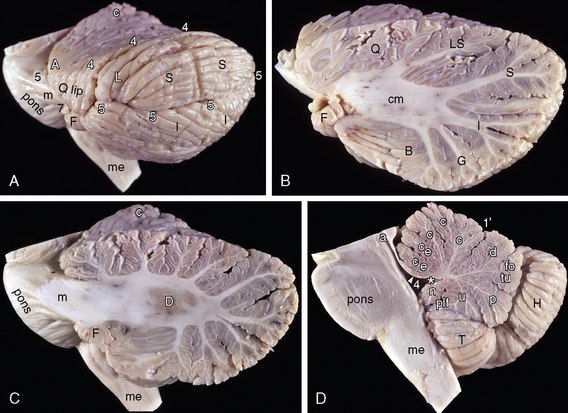
FIGURE 16-3 Lateral surface and sagittal sections of the cerebellum. Formalin-fixed postmortem specimen after removal of the vessels and pia-arachnoid. A, Lateral view. The cerebellum has a characteristic undulant contour. The highest point on the cerebellum is the culmen (c) of the vermis. The anterior angle (A), lateral angle (L), and anterolateral margin are well seen. The posterior superior fissure (4) and the great horizontal fissure (5) define the superior semilunar lobule (S). They meet at the lateral aspect of the anterior surface and angle together toward and then superior to the flocculus (F). The middle cerebellar peduncle (m) passes laterally from the pons (pons) into the cerebellar hemisphere. me, medulla; 5, CN V (trigeminal nerve); 7, CN VII (facial nerve). B, Sagittal section through the flocculus (F) and the lateral portion of the hemisphere shows the central body of cerebellar white matter (corpus medullare) (cm) extending outward into each lobule of the vermis in a characteristic branching pattern. Note the quadrangular lobule (Q), lobulus simplex (LS), superior semilunar lobule (S), inferior semilunar lobule (I), lobulus gracilis (G), and the biventral lobule (B). C, Lateral sagittal section through the middle cerebellar peduncle (m). The peduncle arises from the pons and passes into the corpus medullare. The dentate nucleus (D) lies centrally surrounded by the corpus medullare. D, Midsagittal section through the lowermost midbrain, pons (pons), medulla (me), aqueduct (a), and fourth ventricle (4). Midsagittal section through the cerebellum displays the midline vermis, the posterior cerebellar notch behind the vermis, and the medial surfaces of the opposite tonsil (T) and cerebellar hemisphere (H). The white matter of the vermis has a characteristic, spokewheel pattern designated the arbor vitae. There are nine lobules of the vermis. In order from cranial to caudal: the lingua (arrowhead) is closely applied to the superior medullary velum. The central lobule (ce) shows paired fronds that face the posterior surface of the midbrain and upper pons. The culmen (c) forms the apex of the vermis and typically displays a vertically oriented spike of white matter. In this specimen, the anteriormost part of the culmen more closely resembles a third frond of the central lobule (see Fig, 16-4A). The declive (d), folium (fo), and tuber (tu) appear to arise from a common spike of white matter that is oriented nearly perpendicular to the floor of the fourth ventricle (i.e., the posterior surface of the brain stem). The angle formed by the floor of the fourth ventricle and the central spike normally measures more than 90 degrees (see precise definition in text). The pyramis (p) forms the posteroinferior curvature of the vermis, typically diametrically opposite the apex of the culmen. The midline uvula (u) lies directly atop and between the two tonsils, giving it its name. The nodulus (n) indents the posterior medullary velum, giving it its characteristic shape. The fastigial recess (asterisk) of the fourth ventricle (4) is the midline angle of the fourth ventricle between the superior and the inferior medullary vela. The primary fissure (1′) separates the anterior from the posterior lobes of the cerebellum. The vermian portion of the primary fissure lies between the culmen and the declive. The posterolateral fissure (plf) separates the posterior from the flocculonodular lobes. The vermian portion of the posterolateral fissure separates the uvula from the nodulus. Note that the white matter of the vermis has a “spokewheel” configuration, whereas the folia and fissures of the tonsil (T) are oriented parallel to each other, perpendicular to the posterior surface of the medulla. The choroid plexus passes inferiorly along the floor of the fourth ventricle toward the midline foramen of Magendie.
The superior surface shows three large lobules delimited by three fissures. From anterior to posterior, these are the primary fissure, the posterior superior fissure, and the medial portion of the great horizontal fissure. The anteriormost (primary) fissure is trapezoidal and lies between the culmen/quadrangular lobule in front and the declive/lobulus simplex behind. It separates the anterior from the posterior lobe of the cerebellum and is the deepest fissure in the vermis.2 The next most posterior fissure is the rounder posterior superior (postclival) fissure. It has a rounded shape and just “kisses” the anterior edge of the posterior cerebellar notch in the midline. This posterior superior fissure separates the declive/lobulus simplex anteriorly from the folium/superior semilunar lobule posteriorly. Two small oblique fissures arise in the posterior cerebellar notch and extend posterolaterally for a short distance. These are the small, medial portions of the great horizontal fissures that become very much larger on the posterior surface. The great horizontal fissures separate the folium/superior semilunar lobules anteriorly from the tuber/inferior semilunar lobules posteriorly. On the superior surface, the midline vermis is delimited laterally by two shallow paramedian grooves and by the abrupt angulations made by the cerebellar fissures as they pass from the hemispheres laterally to the vermis medially.
Posterior (Occipital) Surface
The posterior surface is round with an undulant lateral border (see Figs. 16-1C and 16-2C). The superior edge of the posterior surface is the posterolateral margin that is formed by the superior semilunar lobule. This margin is highest in the midline and slopes gently downward as it passes laterally. The inferior edge of the posterior surface is the biventral ridge, formed by the inferior margin of the biventral lobule. The lateral border of the posterior surface is sinuous, convex laterally more superiorly and convex medially most inferiorly. On the posterior surface, the medial border of each hemisphere is largely convex medially. The posterior surfaces of the hemispheres are clearly demarcated from the vermis by deep paravermian grooves/fissures.
Up to seven variably large fissures are seen along the posterior and inferior surfaces of the cerebellum (see Table 16-1). The most superior, deepest, and most prominent fissure on each side is the great horizontal fissure. This starts on the superior surface near the midline, passes inferolaterally onto the posterior surface, and then continues inferolaterally across the entire posterior surface of the hemisphere. It passes approximately 1 cm inferior to the lateral angle of the cerebellum and then swings onto the anterior (petrosal) surface of the cerebellum. The great horizontal fissure separates the folium/superior semilunar lobule above from the tuber/inferior semilunar lobule below (Fig. 16-2).
Inferior to this point, the vermian and hemispheric structures realign with each other and curve onto the undersurface of the cerebellum, like a dog with its tail between its legs. Tucked deeply into the posterior surface (and not readily visible on the posterior surface), the postpyramidal (secondary) fissure separates the pyramis/biventral lobule above from the uvula/tonsils below. The posterolateral fissure separates the uvula/tonsils behind from the nodulus/flocculus in front. The relationships of the pyramis, uvula, and nodulus are best appreciated on midsagittal sections, rather than by surface inspection (see Figs. 16-3B and C). Along the posterior surface, two deep paravermian grooves delimit the lateral borders of the inferior vermis, incompletely separating the vermis from the cerebellar hemispheres lateral to it.
Anterior (Petrous) Surface
The anterior surface of the cerebellum is largely obscured by the brain stem (see Fig. 16-2E). The paired middle cerebellar peduncles arise from the lateral borders of the pons and dive deeply into the cerebellar hemispheres, enclosed by a superior and inferior rim of cerebellum. On each side, the superolateral rim and the upper edge of the anterior surface is the anterolateral margin. The small band of tissue that lies between the anterolateral margin and the brain stem itself is designated the quadrangular lip. The lip is formed, from medial to lateral, by the quadrangular lobule, the lobulus simplex, and the superior semilunar lobule. The band of cerebellum inferior to the middle cerebellar peduncle and lateral to the medulla is composed of the flocculus immediately inferior to the peduncle and (from lateral to medial) by the inferior semilunar, gracile, and biventral lobules more peripherally. On each side, the margin of the hemisphere situated immediately lateral to the medulla is the biventral ridge. The cerebellomedullary fissures separate the biventral ridges from the medulla on each side.
Sectional Anatomy of the Cerebellum
Midline sections of the brain stem and cerebellum (see Figs. 16-3B and D) display the midbrain, pons, and medulla of the brain stem anterior to the aqueduct and fourth ventricle. The roof of the fourth ventricle is formed by the anterior (superior) medullary velum and the posterior (inferior) medullary velum. These meet at a sharp angle called the fastigial recess of the fourth ventricle. Behind the fourth ventricle lies the vermis. The vermis has a variable shape, sometimes trilobed like a three-leaf clover and at other times very smoothly rounded like a “Pacman” character. The nodulus of the vermis indents the fourth ventricle in the midline, along the posterior medullary velum, immediately inferior to the fastigial recess. It is the upper border of the nodulus that creates the curved midline contour of the posterior medullary velum. The paired cerebellar tonsils lie just off the midline to each side. They are ovoid and appear to align parallel with the long axis of the medulla. The upper poles of the tonsils indent the posterior wall of the fourth ventricle just to each side of midline, forming the paired posterior superior recesses of the fourth ventricle. It is the upper poles of the tonsils that create the curved contours of the posterior fourth ventricle laterally on each side. In midline sections, the upper pole of the tonsil is obscured by the midline vermis while the lower pole extends caudal to the vermis. The vermis and the tonsil show very different relations to the fourth ventricle and very different folial patterns. The folia and fissures of the vermis appear to radiate outward from the fastigial recess of the fourth ventricle like a spokewheel. The folia and fissures of the tonsils align perpendicular to the long axis of the medulla.
The three lobes and nine lobules of the cerebellum are readily seen in the vermis. Clockwise, from rostral to caudal the three lobes are the anterior, posterior, and flocculonodular lobes. Clockwise, from rostral to caudal, the nine lobules are the lingula, central lobule, and culmen of the anterior lobe; the declive, folium, tuber, pyramis, and uvula of the posterior lobe; and the nodulus of the flocculonodular lobe. The individual lobules are delimited by well-defined fissures (see Figs. 16-3D and 16-4). The primary fissure separates the anterior from the posterior lobe. The posterolateral fissure separates the posterior from the flocculonodular lobe.
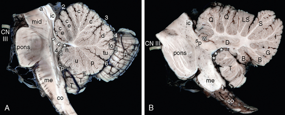
FIGURE 16-4 Midline (A) and parasagittal (B) sections of the cerebellum. Formalin-fixed specimen with vessels and pia-arachnoid intact. A, The lobules of the vermis are labeled as in Figure 16-3D. The fissures associated with these lobules are marked with white numbers: precentral fissure (1), preculminate fissure (2), primary fissure (3), posterior superior fissure (4), great horizontal fissure (5), prepyramidal fissure (6), secondary fissure (7) and posterolateral fissure (8). The posterior superior fissure just “kisses” the depth of the posterior cerebellar notch, so it appears to lie very posterior with respect to the vermis but shows the expected location with respect to the hemisphere (H). This specimen displays the typical configuration of the culmen, with a single near-vertical spike of white matter leading to the apex, posterior to two fronds of the central lobule. The lingua (arrowheads) lies directly upon the superior medullary velum. The nodulus (n) indents the posterior wall of the fourth ventricle in the midline, creating the fastigial recess (asterisk). The orientation of the paramedian perforating vessels to the brain stem is particularly well shown in this specimen. Other labels indicate the inferior colliculus (ic) of the midbrain, the choroid plexus of the fourth ventricle (cp), and the spinal cord (co). B, The upper pole of each tonsil indents the posterior wall of the fourth ventricle, off midline, forming a paramedian, posterosuperior recess of the fourth ventricle. As a result, the posterior surface of the fourth ventricle displays three recesses: a midline fastigial recess that lies more anterior and superior and paired paramedian posterior superior recesses (three asterisks) that lie more posterior and inferior. The choroid plexus (same asterisks) of the fourth ventricle lies along the posterior superior recesses atop the two tonsils. The choroid also extends laterally through the lateral recesses into the cerebellopontine angles and caudally toward or through the midline foramen of Magendie. The dentate nucleus (D) lies within the cerebellar white matter and contributes efferent fibers to the superior cerebellar peduncle (sp). This more lateral sagittal section shows the wing of the central lobule (w), quadrangular lobule (Q), lobulus simplex (LS), superior (S) and inferior (I) semilunar lobules, lobulus gracilis (G), and biventral lobule (B).
The white matter of the vermis forms a prominent branching pattern (the arbor vitae) that appears to radiate out from the fastigial recess of the fourth ventricle like a spokewheel (Fig. 16-4). The cerebellar white matter of each hemisphere extends across the midline, forming a cerebellar commissure. This commissure passes forward into the superior medullary velum. The choroid plexus of the fourth ventricle lies on the intraventricular side of the posterior medullary velum. It is a paired paramedian structure, not a midline structure. On each side the choroid plexus overlies the upper pole of the tonsil and extends both inferomedially toward Magendie and anterolaterally through the lateral recess of the fourth ventricle to emerge into the cerebellopontine angle through the foramen of Luschka.
Serial sections through the cerebellum display this anatomy in detail (Figs. 16-5 and 16-6).
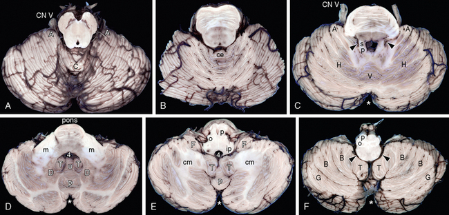
FIGURE 16-5 Serial axial sections through the formalin-fixed cerebellum, displayed from superior to inferior. A, Uncut superior surface displays the typical relationships among the midbrain, culmen of vermis (c), anterior angles of the cerebellum overlying the origins of CN V (trigeminal nerve), array of cerebellar folia co-curvilinear “parallel” with the posterolateral margin and each other, and the course of the blood vessels approximately perpendicular to that folial pattern. B, Section through the pontomesencephalic junction and the upper cerebellum. The folia are continuous from one hemisphere across the vermis to the other side. Their course angles sharply at the paravermian boundary, from oblique to horizontal to oblique again, giving the folia a characteristic trapezoidal configuration. The central lobule (ce) of the vermis faces the tectal plate at this level. This section lies above the posterior cerebellar notch, so the posterior contour of the section is convex. C, Section through the pons, upper fourth ventricle (4), vermis (V), and hemispheres (H). The anterior angles (A) overlie the entries of CNV into the lateral pons on each side. The superior cerebellar peduncles (sp) that form the lateral walls of the upper fourth ventricle appear as thin plates of white matter interposed between the fourth ventricle medially and the ambient cisterns (arrrowheads) laterally. Asterisk indicates the posterior cerebellar notch. D, Section through the midpons and fourth ventricle (4) at level of the upper poles of the tonsils (T). The thick middle cerebellar peduncles (m) pass obliquely from the lateral aspects of the pons into the central white matter (corpus medullare) of each cerebellar hemisphere. The paired dentate nuclei (D) form pleated crescents to each side of the fourth ventricle. The paired tonsils (T) flank the uvula (u) of the vermis. E, Section through the medulla, low fourth ventricle (4), flocculi (F), and pyramis (white p) of the vermis. The medulla shows the characteristic curvatures of the medullary pyramids (black p), olives (o), and inferior cerebellar peduncles (ip). The flocculi (F) form fronds that extend laterally along the anterior surface of the cerebellum. The paired tonsils protrude toward the midline from the medial borders of each hemisphere. The pyramis of the vermis displays its characteristic bulbous quadrifoil shape at the depth of the posterior cerebellar notch (asterisk). This section displays the lowest portion of the cerebellar white matter (cm). A tiny hemorrhage is seen in the right inferior cerebellar peduncle. F, Section through the medulla, tonsils, and biventral lobules inferior to the cerebellar white matter and vermis. The pyramids (p) and olives (o) of the medulla are well shown. The paired tonsils (T) lie half behind and half alongside the medulla. The portions of the tonsils situated behind the medulla are convex medially. The portions situated lateral to the medulla are concave medially. The cerebellomedullary fissures (black arrowheads) separate the tonsils and biventral lobules from the medulla on each side. The folia and fissures of the biventral lobule appear co-curvilinear with the lateral border of the hemispheres. Asterisk indicates posterior cerebellar notch.
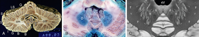
FIGURE 16-6 Cerebellar nuclei. Two formalin-fixed human cerebella. A, Unstained coronal section through the fourth ventricle, corpora medullare (cm) of the two hemispheres, the paired tonsils (T), the nodulus (n), and the uvula (u). The deep cerebellar nuclei (roof nuclei) are arrayed symmetrically around the roof of the fourth ventricle. In order from medial to lateral, these roof nuclei are the paired fastigial (F), globose (G), emboliform (E), and dentate (D) nuclei. In the coronal plane, the surface fissures and the central spikes of white matter for each lobule localize and identify the quadrangular lobule (Q), lobulus simplex (LS), superior (S) and inferior (I) semilunar lobules, lobulus gracilis (G), and biventral lobule (B). B, Perl (ferricyanide) stain of an axial section comparable to the section shown in Figure 16-5D. Blue coloration of the dentate nuclei (D) indicates the presence of ferric (Fe3+) iron. Peripheral blue-green coloration is an artifact of prolonged formalin fixation. C, Composite image of postmortem specimen. Oblique 9.4-T MR image demonstrates the roof nuclei arrayed about the fourth ventricle (4V) within the cerebellar white matter (7). From medial to lateral these are the fastigial (4), globose (3), emboliform (2), and dentate (1) nuclei. C, Cerebellar hemisphere; V, vermis.
(C, Modified from Naidich TP, Duvernoy HM, Delman BN, et al (eds). Duvernoy’s Atlas of the Human Brainstem and Cerebellum. New York, Springer-Verlag, 2009.)
Finer Architecture of the Cerebellum
Cerebellar Cortex
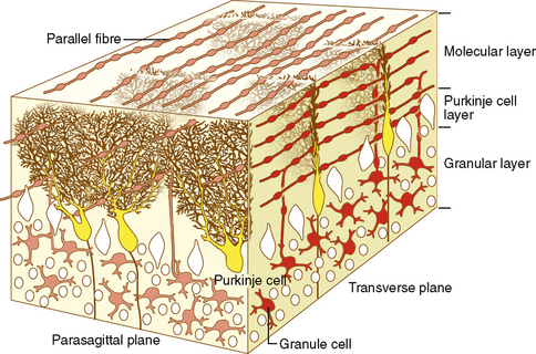
The cortex of the cerebellum is composed of three layers.1 From superficial to deep, these are the superficial cell-poor molecular layer, a middle one-cell-thick Purkinje cell layer, and the deep granule cell layer (Fig. 16-7).1 The Purkinje cell layer gives rise to the efferent, output fibers from the cerebellum. The granule and molecular layers contain processes of the Purkinje cells and multiple interneurons (Fig. 16-8).1
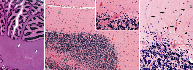
FIGURE 16-7 Cytoarchitecture of the cerebellar cortex (hematoxylin & eosin stain). A, Cerebellar folia (original magnification, ×3). The cerebellar folia resemble the fronds of a fern leaf. The dentate nucleus (white arrows) is seen faintly within the white matter. B, The cerebellar cortex has three layers resting on a white matter (WM) base: the outer cell-sparse molecular layer (M), the middle single-cell-thick Purkinje cell (P) layer, and the inner densely cellular granule cell layer (G) (original magnification, ×100). Inset, Magnified view of the single Purkinje cell indicated by the white arrow (×250). C, Purkinje cells and their processes. The goblet-shaped Purkinje cells (P) extend their dendritic processes (black arrows) outward into the molecular layer (M), perpendicular to the surface of the cerebellum and perpendicular to the parallel fibers (original magnification, ×250). The numerous granule cells (G) lie deep to the Purkinje cell layer.
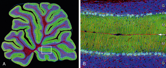
FIGURE 16-8 Histology of the cerebellar cortex: rat brain. Dual-photon multicolored fluorescent laser microscopy. In this specimen, the Purkinje cell neurons have been rendered light blue with green processes, the Bergmann glial cells and their processes red, and DNA-containing cell nuclei blue by use of three differently colored fluorescent dyes (fluorescein, rhodamine, and Hoechst 33342). A, Cross section of the whole cerebellum. The white box indicates the area and orientation of part B. B, Magnified view of two cortical layers apposed across the cerebellar fissure (white arrow). The myriad nuclei within the highly cellular granule cell layer (G) give the granule cell layer a blue coloration. The one-cell-thick Purkinje cell layer (P) stains bluish green. The Purkinje cells send their processes far out into the molecular layer (M).
(Images courtesy of Thomas Deerinck and Mark Ellisman, The National Center for Microscopy and Imaging Research, University of California, San Diego.)
The cerebellar white matter contains afferent mossy fibers, afferent climbing fibers, and Purkinje cell axons en route to the cerebellar/vestibular nuclei.1 The mossy fibers originate in the spinocerebellar tracts ascending from the spinal cord, in the cuneocerebellar tracts ascending from the medulla, and in the vestibular nuclei, reticular nuclei, and pontine nuclei of the brain stem. They terminate on dendrites of granule cells to form islands of synaptic complexes (glomeruli) within the granule cell layer. Climbing fibers originate exclusively from the contralateral olivary complex. They terminate on Purkinje cell dendrites in the molecular layer.
Within the cerebellar white matter, the fibers are highly organized into mutually perpendicular systems formed by long parallel fibers that course parallel to the cerebellar surfaces and perpendicular fibers that are restricted to specific narrow parasagittal compartments (stripes) aligned perpendicular to the cerebellar surfaces (Figs. 16-9 to 16-11).