Case presentation
A 12-month-old female presents following a motor vehicle accident. The child was reportedly secured in a rear car seat; however, the child was found outside of the vehicle lying on the pavement. The two-vehicle crash occurred at a high rate of speed (70–80 miles/hour), and there was considerable damage to both vehicles but no deaths at the scene.
In the Emergency Department, the child has a waxing and waning mental status, with her Glasgow Coma Scale (GCS) score varying between 8 and 12. She has some bruising to the right side of the forehead without crepitus or step-offs and her face seems uninjured; she is in a cervical collar applied by Emergency Medical Services prior to arrival. Examination of the neck shows no abnormalities, but the child seems to not want to move her neck and there appears to be some pain to palpation of cervical vertebrae, although it is hard to determine the exact spinal level. She is not moving her left leg well and there is swelling of the thigh.
Imaging considerations
Plain radiography
Plain cervical spine radiographs should be the initial radiologic study to evaluate for cervical spine injury in most children who require workup. The three-view spine series (cross-table lateral, anterior-posterior [AP], and, when obtainable, open-mouth odontoid) should be obtained as the initial study. Sensitivity for the cross-table lateral view for injury is 79% and increases to 90% with the addition of the AP or odontoid views. It is necessary to review the images to ensure that all of the cervical vertebrae are visualized, as the most frequent cause of a missed injury of the vertebral body is an inadequate film series.
Computed tomography (CT)
The sensitivity and specificity of CT for detecting cervical spine bony injury are ≥98%. However, CT scan should be ordered judiciously, employing the principles of ALARA. Obtaining a cervical spine CT exposes a child to a greater radiation dose than a plain film series does. In 2002, a helical cervical spine CT was shown to expose a child’s skin and thyroid to approximately 10 and 14 times the radiation, respectively, compared to five-view radiographs of the cervical spine. With current newer CT technology, the exposure difference would not be as great but still would be higher with CT than plain radiography. In children with a GCS score of ≤9, neurologic deficit on physical examination, or inadequate cervical spine radiographs (in the setting of a high likelihood of injury based on history and physical examination), imaging with CT of the cervical spine instead of plain radiographs is indicated.
Magnetic resonance imaging (MRI)
In patients with an abnormal neurologic examination, or when imaging of the spinal cord or other soft tissues of the spinal column is necessary, MRI should be obtained. MRI is less sensitive than CT for the detection of bony injuries of the cervical spine and craniocervical junction but is better than CT for the identification of intervertebral disk herniation, ligamentous injuries, and spinal cord injuries as well as soft tissue trauma.
Imaging findings
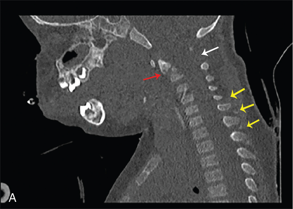
Since the child underwent CT of the head, the cervical spine was imaged with this modality. Selected images of the cervical spine study are provided here. A cervical (C2) spine fracture with increased distance between C1 and C2, spinous process fractures. C1 and C2, and multiple spinous process fractures were demonstrated ( Fig. 48.1 ).