Chemotherapy and hormones

Chapter 19 Chemotherapy and hormones


Introduction


Chemotherapy is the use of cytotoxic (cell poisoning) drugs to control tumour growth. Over the last 30 years, there have been advances in the management of several solid tumours and haematological malignancies, such as testicular teratoma, the leukaemias, childhood cancers and choriocarcinoma. Over 70% of childhood cancers are now curable and the cure rate for teratoma is over 95%. What has been more challenging is the use of cytotoxics in the management of the common tumours, such as non-small lung cancer, breast and bowel cancer. These tumours often present in the metastatic stage and are relatively resistant to chemotherapy. Despite intense research, chemotherapy for these tumours remains palliative in intent rather than curative. One of the major drawbacks of cytotoxic agents is that they affect all rapidly dividing cells, and do not discriminate between normal tissues and tumours, hence, the toxicity of chemotherapy. We are now entering an exciting era in oncology with new targeted therapies becoming available. These novel agents are designed to exploit our increased understanding of molecular oncology and will hopefully be more successful in overcoming drug resistance and reducing the side effects of treatment.


Hormone therapy is another systemic treatment for cancer. It may involve inhibiting the production of endogenous hormones or introducing synthetic ones. There are now many different hormone preparations available and they play a vital role in the treatment of tumours such as prostate and breast cancer.


Principles of cytotoxic therapy


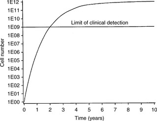
To understand the rationale of cytotoxic chemotherapy it is important to recognize the features of tumour growth. The asymmetric sigmoidal growth curve, the ‘Gompertzian growth curve’ (Figure 19.1) describes the natural history of tumour growth. By the time that a tumour is clinically detectable, the majority of its growth has already occurred. In the early exponential phase of growth, the rates of tumour cell growth and tumour cell loss are proportional to the tumour cell burden at any point. Since most anticancer agents are more toxic to proliferating cells and most tumours are in a relatively slow phase of growth when diagnosed (i.e. they lie high and towards the plateau of the Gompertzian growth curve), it explains the limited effectiveness of chemotherapy for many cancers. The reason for tumour cytoreduction (e.g. by surgery) before chemotherapy is to bring the tumour to a lower point on the growth curve when the growth fraction of the tumour rises. The concept of moving the tumour down the Gompertzian curve underpins the rationale of adjuvant chemotherapy.


Unfortunately, it is not only the proliferating cells that must be eradicated by chemotherapy but also the small population of clonogenic cells mainly in G0 phase. This explains some of the inherent problems of tumour chemoresistance. Cytotoxic drugs prevent cell division by inhibiting DNA replication. Unfortunately, these agents are not specifically acting against malignant cells, and damage both normal and malignant proliferating cells. A careful balance has to be kept between toxicity to the tumour and to the patient’s normal tissues. What distinguishes normal and malignant cells is the failure of the malignant cell, unlike normal cells, to recover from cytotoxic damage. It is exploitation of these differences that underpins the role of targeted therapies.


Drug resistance


A variety of host factors influence the response to chemotherapy, these include the growth fraction of the tumour, the availability of the drug to the tumour and drug resistance. Resistance to chemotherapy may be intrinsic or acquired. Some tumours are intrinsically chemoresistant and show no response to treatment de novo. In other tumours, there is an initial response followed by relapse due to acquired resistance. Acquired resistance may have a variety of mechanisms. These include:


The multiple drug resistance gene (MDR1) encodes P-glycoprotein. The latter is a membrane-associated efflux pump that is widely found in normal cells and serves to protect them from drug-induced damage. Normally, P-glycoprotein is found in very low levels, however, cancer cells can overexpress MDR1 so conferring resistance to a variety of chemotherapeutic agents. In addition, p53, the ‘guardian’ of the genome and an important mediator of apoptosis (programmed cell death) may be mutated and give rise to chemoresistance in a number of solid tumours.


The reasons for drug resistance are not fully understood. It is common to find that a tumour responds to a particular drug or combination of drugs for a period of time and then ceases to do so. It is thought that within many tumour populations there are genetically determined drug-resistant cells. When the chemosensitive cells have been killed, the resistant population may proliferate. Drug resistance to repeated exposure to a single agent will usually result in cross-resistance to other compounds of the same class of drugs. This is probably due to common transport mechanisms and pathways of metabolism and intracellular cytotoxic targets. However, cancer cells that have become resistant to one class of drugs may retain sensitivity to another class of drugs. Most drugs have a variety of mechanisms of drug resistance.


Some drugs which show excellent cell kill in vitro fail to do so in vivo. There may be multiple reasons for this. For example, if the tumour is in a sanctuary site, such as the central nervous system (CNS), the drug does not cross the blood–brain barrier and is therefore ineffective. There is also evidence that some tumours exhibit drug resistance that is partly due to host factors which modify the pharmacokinetics of the anticancer agent in vivo. Chemotherapy is most effective in killing proliferating cells. While the growth fraction is high in many chemosensitive tumours, such as the lymphomas and testicular teratomas, it is relatively low in many common tumours, e.g. colorectal cancer. Finally, in parts of the tumour the blood supply tends to be poor. This not only results in an inadequate concentration of drug reaching the tumour, but also the hypoxia reduces the growth fraction.


Selection and scheduling of chemotherapy agents


In an attempt to improve the curative potential of chemotherapy, agents with proven anticancer properties against a particular tumour but with different mechanisms of action and, as far as possible, non-overlapping toxicities are combined. This is known as combination chemotherapy. For example, in treating breast cancer, three agents – cyclophosphamide, methotrexate and 5-fluorouracil (known as CMF) – all have activity against breast cancer as single agents. However, their response rate as a combination (around 40%) is two- to threefold that of their response rates as single agents. Thus, the overall response is at least additive if not synergistic.


Most schedules of chemotherapy administer the drugs on an intermittent basis to take advantage of the growth kinetics of malignant cells and normal tissues. After each pulse, the normal and malignant cell populations decline due to killing of cells in mitosis. The lowest level of the blood count is known as the nadir. However, whereas the bone marrow recovers to its previous level, the malignant cell population does not. With each subsequent course, this difference is accentuated. If the interval between pulses is too short, toxicity may prevent the delivery of further pulses on schedule, conversely, if the interval is too long, the tumour may regrow between courses. The total dose that can be administered is limited by the tolerance of normal tissue. Toxicity is often cumulative and may be irreversible. For example, the major dose-limiting toxicity of the anthracycline, doxorubicin is cardiotoxicity.


Route of administration


The route of administration is governed by the solubility, chemical stability and local irritant properties of the agent. The simplest route, and the one that patients prefer, is the oral route. Patients can take their tablets at home with intermittent outpatient visits to monitor treatment. Unfortunately, many cytotoxic drugs are unstable and inactivated in the stomach, rendering them ineffective. Intravenous injection is the commonest route of administration of cytotoxic agents since it gives direct access to the systemic circulation. It can be done by delivery of a bolus dose or by infusion. Continuous infusions can be given (e.g. 5-fluorouracil) linked to a battery-operated pump worn around the patient’s waist. The risks of intravenous administration are the introduction of infection and damage to the tissues around the site of administration if extravasation occurs.


It is possible to administer intraperitoneal chemotherapy, for example, in ovarian cancer. However, the drug absorption is variable and there are concerns about adverse effects, such as the development of adhesions. Intra-arterial administration has the advantage of delivering the drug in high concentration to the tissue supplied by the artery. Its limitations are the complexity of administration and the difficulty in correctly identifying the arterial supply of the tumour. Its main use is in the infusion of chemotherapeutic agents into the hepatic artery of patients with liver metastases from colorectal cancer. Intrathecal injection is used to deliver drugs in high dose into the CNS. Many cytotoxic drugs do not cross the blood–brain barrier and are therefore unable to kill tumour cells within the CNS. Methotrexate is the agent most commonly given by this route, for example, when the meninges are involved in lymphoma.


Side effects of chemotherapy


The main normal tissues damaged by cytotoxic therapy are those with rapidly dividing cell populations: the bone marrow, the gastrointestinal epithelium, the hair and the germ cells of the testis. By contrast, there is little effect on non-proliferating tissues, such as skeletal muscle and nervous tissue. The most common side effects of individual cytotoxic drugs are shown in Table 19.1. The most dramatic improvement in the management of side effects was with the development of the 5-hydroxytryptamine antagonist antiemetics. More recently the Neurokinin-1 antagonist aprepitant has improved the emesis of patients undergoing cisplatin based chemotherapy. Typical antiemetic regimens are shown in Table 19.2. Other drugs that have improved drug delivery have been the recombinant growth factors for red (erythropoietin) and white cells (human granulocyte colony-stimulating factor (G-CSF)). These agents stimulate the release of progenitor cells from the bone marrow and are useful in the treatment and prevention of anaemia and neutropenia.

Mar 7, 2016 | Posted by in GENERAL RADIOLOGY | Comments Off on Chemotherapy and hormones

Full access? Get Clinical Tree

Get Clinical Tree app for offline access