32.1 Soft tissue windows: heart
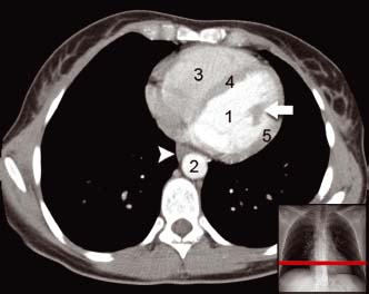
Intravenous contrast is seen in the left ventricle (1) and descending aorta (2). Structures of the heart such as the right ventricle (3), intraventricular septum (4), left ventricular free wall (5) and papillary muscles (arrow) are clearly seen. The tissue adjacent to the aorta is the oesophagus (arrowheads)
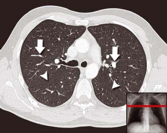
Here the major fissures can be seen on both sides (arrowheads). The branches of the pulmonary vessels are seen (arrows) which make up the lung markings on a plain CXR
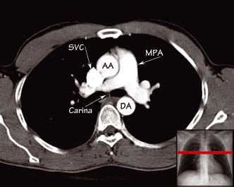
At the level of the carina the main pulmonary artery (MPA) is seen to branch into left and right main pulmonary arteries. The superior vena cava (SVC) lies immediately above the right atrium. The ascending aorta (AA) arches over the pulmonary vessels and major bronchi to join the descending aorta (DA)
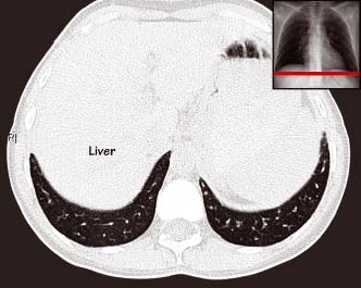
Crescents of lung tissue are seen within the posterior recess of the chest cavity. These parts of the lungs are not easily visible on plain CXR. Soft tissues of the abdomen, such as the liver, are visible immediately below the diaphragm but no detail is provided when viewing with lung window settings
Chest anatomy seen on CT
Related posts:


Stay updated, free articles. Join our Telegram channel

Full access? Get Clinical Tree


