34.1 Primary lung cancer (soft tissue windows)
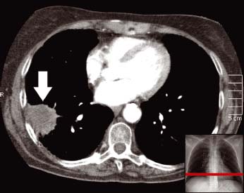
A large spiculated mass is seen adjacent to the pleura in the posterior right lung (arrow). This was biopsied under CT guidance and shown to be a squamous cell carcinoma
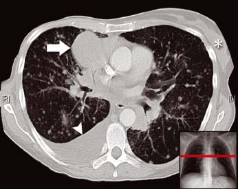
This patient had a known history of breast cancer. Note the right breast is absent following mastectomy. The normal left breast tissue is marked (*). There are multiple small lung nodules that are metastatic deposits. There is also a large round mass next to the right side of the heart (arrow) and a pleural effusion (arrowhead) seen posteriorly in the chest on the right
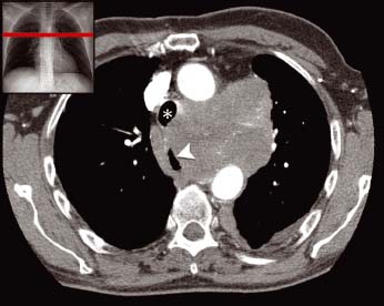
There is a large confluent mediastinal lymph node mass. This is located between the aortic arch and the pulmonary artery (not shown) in a space known as the aorto-pulmonary window. The mass was found to be a lung cancer and is causing deviation of the trachea (*) and oesophagus (arrowhead) to the right
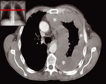
The left lung is encased in grossly thickened pleura (*). This patient has end-stage mesothelioma. The tumour mass has started to deviate the mediastinal structures towards the other side of the chest
Lung Cancer (see Chapter 14)
A lung cancer may be suspected from appearances on CXR (e.g. a focal mass or lobar collapse) or from the clinical history (e.g. cough and weight loss in a smoker). However, further imaging with CT is required for TNM (tumour, nodes, metastases) staging and for planning of procedures such as biopsy and surgery.
- Staging of lung cancer is determined by the TNM status.
- T – Tumour size, position and invasion of adjacent structures.
- N – Lymph node number and position.
- M – Metastatic disease. Common sites for metastasis include the adrenal glands and liver. Other sites may also be imaged depending on the clinical suspicion (e.g. CT of the brain or NM bone scan).
- Biopsy planning
Related posts:

Stay updated, free articles. Join our Telegram channel


Full access? Get Clinical Tree


