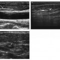
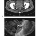
4 Chest Mass Biopsy The first percutaneous transthoracic needle biopsy was reported in 1883; a century later, ultrasound guidance was used to biopsy a lung nodule. It is now common practice and has become an invaluable diagnostic procedure to determine the etiology of masses arising from the lung, pleura, mediastinum, and the chest wall. As image resolution improves, this technique is becoming more accurate, safer, and more widely accepted. Ultrasound has several strengths as a biopsy guidance system. It is readily available, relatively inexpensive, and portable. In addition, it uses no ionizing radiation and can provide guidance in multiple transverse, longitudinal, or oblique planes. The greatest advantage, however, is that it allows real-time visualization of the needle tip as it passes through tissues into the target. This allows precise needle placement and avoidance of important intervening structures. In addition, color flow Doppler imaging can help prevent complications of needle placement by identifying the vascular nature of a mass and by allowing the clinician to avoid vascular structures lying within the needle path. Air in the lungs intervening between the transducer and the lesion prevents its use in the majority of pulmonary lesions. However, when a pulmonary process abuts the pleura or has arisen within the pleura, the chest wall, or a portion of the mediastinum contiguous with the chest wall, ultrasound may be the method of choice for localizing the lesion and guiding the biopsy needle. This may be especially true for apical pulmonary and extrapulmonary lesions for which computed tomography (CT) and fluoroscopy cannot demonstrate an easy access route. Ultrasound-guided biopsy is significantly faster than CT-guided biopsy with average procedure times of 31.4 minutes and 45.2 minutes, respectively. The diagnostic yield with ultrasound guidance is also superior to CT with accuracies of 91% and 71%, respectively. The overall accuracy of ultrasound-guided biopsy ranges between 91 and 98%. Ultrasound-guided needle biopsy can be classified by the preferred method of approach and anatomical location of the lesion. There are no absolute contraindications to percutaneous chest biopsy. In some patients, the platelet count may be normal, but platelet function is decreased in uremia, liver disease, hematologic neoplasms, and in patients taking acetylsalicylic acid. Prior to the procedure, the following should be rechecked: Hospitals have their own policies for obtaining verbal or written informed consents. Regardless of the policy, the procedure, its inherent risks, alternatives, and benefits should be explained in a manner that the patient understands. The physician must be empathetic of patient apprehension regarding pain, and reassurance helps gain patient cooperation during the procedure. An apprehensive patient will not follow breathing instructions and will cause unnecessary delays with poor biopsy results. The patient need not be fasting and no blood is required for transfusion. Therefore, the procedure can be performed on an outpatient basis. Premedication is only advised for percutaneous biopsies of mediastinal lesions. Conscious sedation is induced using midazolam and fentanyl citrate in addition to administration of local anesthesia. For mediastinal biopsies, it is also advised to have noninvasive blood pressure monitoring with continuous pulse oxymetry. The choice of transducer for biopsy is important. A 2—5 MHz convex probe allows visualization of deeper structures; a sector probe with a small footprint allows a wider field of view and is also convenient to place in the intercostal space and suprasternal notch. For superficial lesions a higher frequency linear probe of 5—10 MHz will improve resolution and depict relationship of the lesion to the pleura. When necessary, color Doppler is used to detect potential blood vessels in the needle path. Doppler sensitivity should be set to a low velocity (typically 0.25 m/s) and the wall filter is adjusted to minimize rejection of small frequency shifts to avoid interference from respiratory and cardiac movements. The patient is positioned depending upon the chosen needle entry site. For a parasternal, transsternal, and anterior wall approach, the patient should lie supine. For supraclavicular and suprasternal approach, a pillow is placed under the shoulders and the neck hyperextended. If the approach is lateral, the patient can be positioned in supine, oblique, or lateral decubitus position. The posterior chest is best imaged with the patient sitting upright. Raising the arm above the patient’s head increases the rib space on the ipsilateral side thereby facilitating scanning and allowing for a larger window for needle entry. Percutaneous needle biopsy (PNB) can be performed by the patient’s bedside or in the department. It is useful to have the following three probes at hand: As with other ultrasound-guided biopsies, there are two methods for acquiring tissue, fine-needle aspiration biopsy (FNAB) and core needle biopsy (CNB). FNA can be used for sampling both solid (soft tissue) and fluid. A fine needle is defined as one with an outside diameter of <1 mm and is generally 20—25 gauge in size. FNAB causes less pleural and pulmonary trauma than cutting needles used for CNB. Aspiration produces a highly diagnostic sample for cytologic analysis and is particularly useful for small mediastinal lesions that make the placement and activation of cutting needles (CNB) a daunting prospect. However, the cell aspirate from FNAB may not be sufficient for a definitive diagnosis, thereby necessitating a CNB.
Classification
Indications
Contraindications
Absolute Contraindications
Relative Contraindications
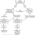
Preprocedural Evaluation and Preparation of the Patient
Equipment
Percutaneous Needle Biopsy
Fine-Needle Aspiration Biopsy
Related posts:
Stay updated, free articles. Join our Telegram channel

Full access? Get Clinical Tree


