• Occipitocervical cephalocele containing cerebellum ± brainstem in conjunction with C1-C2 spina bifida = Chiari 3 malformation
• Distinct malformation; not just a Chiari 2 malformation with encephalocele
(Left) Sagittal T2WI MR reveals a large defect in the ventral chondral portion of the supraoccipital bone and opisthion of the foramen magnum. Gliotic cerebellar tissue protrudes into a large sac. Note displacement of the brainstem and basilar artery .
(Right) Sagittal T1WI MR shows a large meningoencephalocele composed of meninges, CSF, cerebellum , brainstem , and upper cervical spinal cord herniated through a bone defect in the lower occiput and upper cervical spine.
(Left) Bone CT 3D shaded surface rendering of the posterior calvarium/upper cervical spine in a Chiari 3 malformation (CM3) patient shows a large defect of the ventral chondral and squamous supraoccipital bones, in conjunction with upper cervical spina bifida .
(Right) Sagittal MRV demonstrates typical venous abnormalities of Chiari 3. The straight sinus and vein of Galen is severely hypoplastic. Large occipital sinuses , rather than transverse sinuses, are present.
TERMINOLOGY
Abbreviations
• Chiari 3 malformation (CM3)
Synonyms
• Chiari III, rhombencephalocele
Definitions
• Combined cephalocele + myelocele herniating through high cervical ± low occipital dysraphic defect


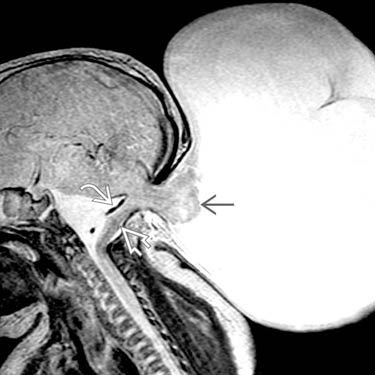
 protrudes into a large sac. Note displacement of the brainstem
  and basilar artery
  .
.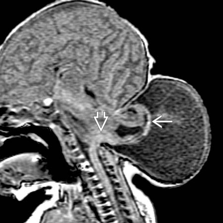
 , brainstem
  , and upper cervical spinal cord herniated through a bone defect in the lower occiput and upper cervical spine.
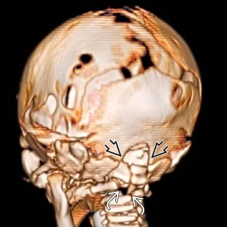
 of the ventral chondral and squamous supraoccipital bones, in conjunction with upper cervical spina bifida
  .
.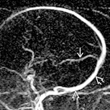
 and vein of Galen is severely hypoplastic. Large occipital sinuses
  , rather than transverse sinuses, are present.