Location: Epiphysis or apophysis of long bones (proximal femur and humerus), flat, and short bones.
Clinical: Modest pain.
Imaging: On x-ray, like a chondroblastoma. Osteolytic lesion with small calcifications, sharp margins, irregular, and ill-defined sclerotic rim. On CT, spots or small granules typical of the cartilaginous tumors.
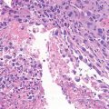
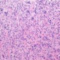
Histopathology: Lobular tissue with clear cells: central nucleus, extremely vacuolated cytoplasm, strongly PAS positive. Rare mitotic figures. Peripheral reactive giant cells, possible areas of well-differentiated chondrosarcoma, and intercellular calcification like chondroblastoma, cystic spaces, and reactive osteoid.
Course and staging: Slow growth, recurrence is possible when intralesional margins are obtained, metastases are exceptional. Usually, stage IA.
Treatment: Wide resection. Prognosis is good.
Key Points
Clinical | Epiphysis and apophysis in adults |
Radiological | Osteolytic, like a chondroblastoma |
Histological | Lobules of clear cells, possible areas of hyaline cartilage, giant cells, calcifications, reactive osteoid
Related posts:Stay updated, free articles. Join our Telegram channel
Full access? Get Clinical Tree
 Get Clinical Tree app for offline access

|