Jean Noel Buy1 and Michel Ghossain2
(1)
Service Radiologie, Hopital Hotel-Dieu, Paris, France
(2)
Department of Radiology, Hotel Dieu de France, Beirut, Lebanon
19.1 Three Main Types
19.1.1 Agenesis
19.1.3 Septated Uterus
19.2 Associated Lesions
19.2.2 Endometriosis (Fig. )
19.3 Other Abnormalities
19.3.2 DES-Related Disorders []
19.3.3 Localized Atresia
19.3.4 Vaginal Septum
19.3.5 Imperforated Hymen
19.3.6 Arcuate Uterus
Abstract
The three main types of müllerian duct abnormalities (congenital abnormality of the paramesonephric duct) are agenesis, bicornuate uterus, and septated uterus.
19.1 Three Main Types
The three main types of müllerian duct abnormalities (congenital abnormality of the paramesonephric duct) are agenesis, bicornuate uterus, and septated uterus.
19.1.1 Agenesis
19.1.1.1 Bilateral: Mayer-Rokitansky-Kuster-Hauser Syndrome (Fig. 19.1)


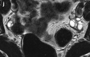
Fig. 19.1
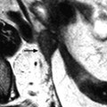
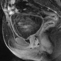
Mayer-Rokitansky-Kuster-Hauser syndrome. Twenty-year-old woman with amenorrhea. (1) Absence of vagina. (a) MR sagittal T2 on the midline displays absence of vagina almost until the introitus, with adipose tissue between two hypointense lines very likely representing the rectovaginal septum (long horizontal arrow) and the vesicovaginal septum (short horizontal arrow). The lower vagina (LV) is present with a cone shape and an apex (oblique arrow) representing the junction with the remnant upper two-thirds vagina. (b–c). Axial T2 at two levels. Through the middle part of the posterior wall of the bladder (b) allows to definitely confirming the absence of vagina until the introitus; At the level of the posterior and inferior part of the pubis (c) displays a normal vestibular vagina. (2) Uterine and tubal remnants. (d, e). Sagittal T2 through the midline (d) displays between the upper part of the bladder and rectum low intensity tissue (arrow). Axial T2 through this tissular mass (e) shows a central mass (central arrow) with irregular borders related to a remnant of the uterus and laterally two tubular structures with a hyperintense to intermediate signal related to tubal remnants (lateral arrow). (f, i). DMR without injection (f) at the arterial (g), venous (h), and late (i) phases. At the arterial phase (g), tubal arterial vessels are displayed in the tubal remnants. At the venous phase (h) and even better on the delayed (i), a significant contrast uptake is visualized in the uterus and tubes remnants, with a characteristic fold appearance in the right tube at the venous phase. (3) Normal ovaries are visualized on axial T2 (j)
Definition
The main features and associated findings of Mayer-Rokitansky-Kuster-Hauser syndrome are summarized in Table 19.1.
Table 19.1
Mayer-Rokitansky-Kuster-Hauser syndrome
Main features |
–46XX chromosome |
–Normal ovaries |
–Hypoplasia of the tubes, aplasia of uterus and vagina until the hymen |
–Normal external female genitalia |
Associated abnormalities |
–Renal anomalies (40–50 % of cases) |
–Associated skeletal or spinal anomalies in 10–12 % of cases |
–Hearing loss in 10–25 % of cases |
Differential Diagnosis
The main differential diagnosis is androgen insensitivity syndrome (AIS). AIS findings are displayed in Table 19.2 (Fig. 19.2).


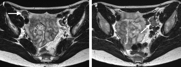

Table 19.2
Findings of androgen insensitivity syndrome (AIS)
–46XY chromosome |
–Testis in the abdomen or in the inguinal canals |
–Aplasia of tubes, uterus, and vagina until the hymen due to secretion of müllerian-inhibiting substance (MIS) also called anti-mullerian hormone (AMH) by the Sertoli cells of the testis |
–Normal external female genitalia (insensitivity to androgens secreted by the interstitial cells of the testis) |


Fig. 19.2
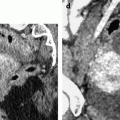
Androgen insensitivity syndrome (AIS). Sixteen-year-old girl with primary amenorrhea. (1) Absence of vagina. (a, b) Sagittal T2W image (a) and axial T2W image at the level of the middle third of the vagina (b) show absence of uterus and collapsed walls of the upper two-thirds of the vagina with no vaginal mucosa seen at this level. Horizontal arrow in (a) points to mucosa of the lower third of the vagina, which is hypoplastic compared to the corresponding part in the Rokitansky syndrome (see Fig. 19.1). (2) Absence of tubal remnants. On sagittal T2 (a), a hypointense tissue (oblique arrow) is displayed in front of the anterior part of the rectum. (c–e) DMR at the level of this tissue before injection (c), at the arterial (d), and late (e) phases. At the arterial phase, no tubal arteries are displayed. Contrast uptake at the venous phase and especially on the delayed (e) is visualized in this little solid tissue (arrow), which is smaller and less well delineated than in Rokitansky syndrome. An associated anterior left cyst is visualized in contact at a higher level with the medial border of the testis, probably related to a cyst of the epididymis. (3) Testis. (f, g) Axial T2W images at the level of the right gonad (arrow in f) and the left gonad (arrow in g), show absence of follicle with a pattern more suggestive of testicles than ovaries. (h–j) DMR at the level of (g), before injection (h), at the arterial (i), and late (j) phases confirms absence of follicles or nodular enhancement in the left gonad. Similar findings were found in the right gonad
Persons with partial AIS exhibit some masculinization at birth, such as ambiguous external genitalia and may have an enlarged clitoris.
19.1.1.2 Unilateral Agenesis
Complete: Unicornis (Figs. 19.3 and 19.4)

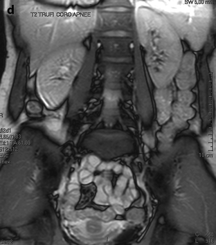
Fig. 19.3
Unicornuate uterus. (a) Sagittal EVS displays a uterus that is deviated to the right (cursors = endometrium). (b) 3D reformation with 2D coronal reconstruction confirms the presence of only one cavity. (c) MR axial T2 confirms the US impression and better displays a banana-like uterus very suggestive of a unicornuate uterus. The mesometrium is well displayed on the right (horizontal arrow), while on the left, it is absent with presence only of the Mackenrodt (paracervix) ligament (oblique arrow). The left round ligament (which originate from the gubernaculums) is visualized in the inguinal canal (vertical arrow). (d) Coronal T2 shows the presence of two normal kidneys. Coelioscopy confirmed the diagnosis
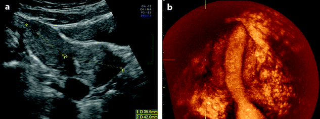
Fig. 19.4
Unicornis right hemiuterus. (a) Transabdominal sonography displays a right banana shaped unicornis hemiuterus measured using two distances. (b) 3D with 2D reformation well displayed the banana shaped endometrium. Coelioscopy confirmed the diagnosis
Definition
Small elliptical uterus deviated on one side of the pelvis with single cornu, with an elliptic endometrial cavity well displayed on US with 3D reformation.
On the side of the absent hemiuterus, the round ligament seems to start directly from the ovary (Fig. 19.5).


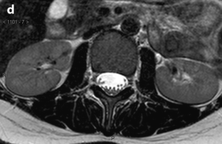


Fig. 19.5
Unicornis. Thirty-four-year-old woman. Infertility. Hysteroscopy: only the left tubal orifice is visualized. MR axial T2 (a) and coronal T2 (b1 and b2) display a left unicornis uterus associated with a right mullerian remnant (arrow). On axial T2, c1 the right round ligament starts from the anterior portion of the right ovary (arrows) and is followed before (c2) (arrow), and during its course through the inguinal canal (c3) (arrow). Although coelioscopy has not yet been performed, this relationship of the round ligament suggests the possibility that in the absence of one uterus cornua, the gubernaculum on the same side (because of the absence of utero-ovarian ligament) starts directly from the ovary. Axial T2 shows (d) two normal kidneys in normal situation
Differentiation from a normal uterus deviated to one side is made on the following findings:
1.
Shape of the uterine fundus is normal with a triangular aspect of the endometrial cavity.
2.
Both round ligaments start from the uterine cornua.
Incomplete: Pseudounicornis
Definition
Unicornis with contralateral rudimentary uterus:
Contralateral rudimentary uterus with endometrium (Fig. 19.6) or without a cavity covered by endometrium.Get Clinical Tree app for offline access


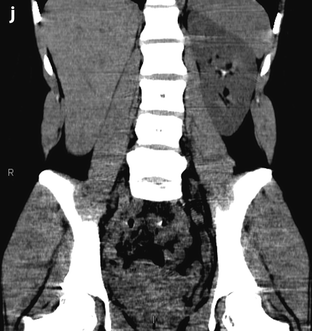
Fig. 19.6
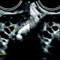
Noncommunicating pseudounicornis uterus with menstrual retention subperitoneal endometriosis and agenesis of the right kidney. Twenty-three-year-old woman with dysmenorrhea. (a) TAS, transverse section, displays a left hemiuterus and a smaller right hemiuterus with medial to it an echogenic collection (arrow). (b) EVS, longitudinal section, depicts a normal left uterus with a thin endometrium. (c) EVS longitudinal section on the right shows a right uterus not communicating with the cervix; a hematic collection of 9 mm thickness is displayed in the endometrial cavity. The endometrium is thin. (d) EVS, through the right collection identified it as a 59 × 23 mm hematosalpinx (cursors). A diagnosis of pseudounicornis uterus is diagnosed. Abdominal ultrasound (not shown) demonstrates absence of the right kidney and normal left kidney. (e, f) MR coronal T2 (e) and axial T2 (f): left hemiuterus and tube are normal. On the right side, a small right hemiuterus is present, with hypointense blood in the endometrial cavity (arrow), without any communication with the left hemiuterus associated with a right hematosalpinx (letters HS). Hypointense thickening of the torus and uterosacral ligament is seen related to endometriosis. Vagina and cervix are unique. (g–i) Axial T1 (g) and T1 fat suppression (h, i
Stay updated, free articles. Join our Telegram channel

Full access? Get Clinical Tree


