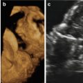
Fig. 14.1
Fetal T2-weighted MRI at 34.6 weeks’ gestation in the three planes (sagittal plane (a), coronal plane (b), axial plane (c)) demonstrates the partial exophytic hyperintense soft tissue mass infiltrating the right orofacial region from the temporaral area to the chin, with invasion of the entire tongue, the floor of the mouth, the right masticator space, and the cheek. Note, in (a), the macroglossia, with the tongue protruding out of the mouth, and partial obstruction of the oral cavity. Note the mass effect of the lesion on the soft palate and compression of rhino-oropharynx (thick arrow) with visualization of a patent and nondistended cervical trachea and larynx (thin arrow in a and c)
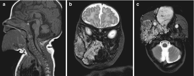
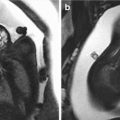
Fig. 14.2
Postnatal MRI at 45 days of life confirmed the voluminous, subcutaneous venous-lymphatic orofacial malformation. T1-weighted sagittal image (a) shows the homogeneous hypointense lesion with corresponding heterogeneous hyperintense signal on T2-weighted images (b, coronal image; c axial image)
A protective tracheostomy was performed at 4 months of age followed by a subtotal glossectomy and multiple laser treatment and foam sclerotherapy.
14.3 Discussion
Subcutaneous mixed venous-lymphatic malformations are usually seen as proliferative tumors such as hemangiomas and/or lymphangioma [1]. Isolated macroglossia is rarely detected prenatally, and even when associated with Beckwith-Wiedemann syndrome, the diagnosis is typically made after birth [4]. In the case reported here, accurate prenatal diagnosis, by means of ultrasound and fetal MRI, of a subcutaneous mixed venous-lymphatic malformation associated with macroglossia aided the genetic counseling and the parents’ decision-making process and allowed appropriate antenatal and post-natal care management [5]. In addition, adding fetal MRI to ultrasound and Doppler study of the fetal vascular anomalies enhances the characterization of these lesions and the accuracy of the antenatal diagnosis and assists with patient selection for an EXIT procedure [6, 7].
Related posts:
 The Genetics of Facial Cleft
 Atypical Facial Cleft Detected by Prenatal Scan and Confirmed by Postmortem Reconstructive Computed Tomography (CT REC)
 Acromelic Frontonasal Dysplasia (Median Cleft Face Syndrome)
 The Fetal Brain in Fetuses with Orofacial Abnormalities
 Magnetic Resonance Imaging (MRI) in the Evaluation of the Fetal Face
 The Role of 2D/3D/4D Ultrasound in the Prenatal Assessment of Cleft Lip and Palate
Stay updated, free articles. Join our Telegram channel

Full access? Get Clinical Tree


