Chapter 76 Conotruncal anomalies are a group of congenital heart defects involving the outflow tract of the heart and great vessels. The conotruncal anomalies include tetralogy of Fallot (TOF), transposition of the great arteries (TGA), double-outlet ventricles, and truncus arteriosus. Interrupted aortic arch type B also is a conotruncal anomaly that will be discussed with other aortic arch anomalies (see Chapter 75). Conotruncal abnormalities are the result of abnormal division or rotation of the primitive truncus during embryologic development.1,2 The common outlet of the embryonic univentricular heart normally undergoes a complex sequence of events to separate into the right ventricular outflow tract (RVOT) and left ventricular outflow tract (LVOT), the aorta, and the main pulmonary artery.3 Control by numerous genes and migration of the mesenchymal cells from the embryonic neural crest are required for this development.1 Mutations in a number of genes have been associated with conotruncal anomalies in humans and animal models.4 Overview: TOF is the most common cyanotic congenital heart defect. The median described incidence of TOF is 356 per 1 million live births in the United States.5 The classic manifestations include RVOT obstruction, ventricular septal defect (VSD), overriding of the aortic root above the VSD, and right ventricular (RV) hypertrophy (Fig. 76-1, A). These findings are a result of underdevelopment of the subpulmonary infundibulum,6 which is associated with anterior deviation of the conal septum. Rather than sitting between the anterior and posterior limbs of the trabecula septomarginalis (a Y-shaped bundle of muscle along the right side of the ventricular septum), the conal septum in TOF typically is fused with the anterior limb, bringing the aorta over the ventricular septum and leading to the malalignment VSD.7,8 The septal malalignment and hypertrophy of the trabeculations of the infundibular free wall result in RVOT obstruction (Fig. 76-1, B). The VSD in TOF is located between the malaligned conal septum superiorly and the muscular septum inferiorly (i.e., a conoventricular septal defect),9 and it typically is large, nonrestrictive, and subaortic. For further discussion of the trabecula septomarginalis, see the section of this chapter on double-outlet RV. Figure 76-1 Three-dimensional steady-state free-precession imaging in an 18-year-old man with unrepaired tetralogy of Fallot. The anatomic appearance of TOF varies, including TOF with pulmonary atresia and TOF with dysplastic (absent) pulmonary valve syndrome. The extent of RVOT obstruction is variable, ranging from minimal obstruction to pulmonary atresia.10 The pulmonary valve often is thickened or fused with doming leaflets and a variably hypoplastic annulus causing valvular stenosis. The size of the main and branch pulmonary arteries also varies. Patients with pulmonary valve atresia have no antegrade flow supplying the pulmonary arteries; instead, pulmonary blood flow is supplied by a patent ductus arteriosus, aortopulmonary collateral arteries, or both (e-Fig. 76-2). The central pulmonary arteries can be absent, discontinuous, or diminutive. In persons with TOF and a dysplastic pulmonary valve, congenital severe pulmonary regurgitation occurs, which often is associated with severe dilatation of the central pulmonary arteries and resultant airway compression (e-Fig. 76-3, A and B).11 e-Figure 76-2 A 2-month-old girl with tetralogy of Fallot and pulmonary atresia. e-Figure 76-3 Turbo spin echo black blood imaging in a 5-year-old boy with tetralogy of Fallot and an absent pulmonary valve after repair with a transannular patch. Etiology, Pathophysiology, and Clinical Presentation: Genetic abnormalities such as chromosome 22q11 deletion, which also leads to DiGeorge or velocardiofacial syndrome, may play an important role in some patients with this disease.12 Many cases are sporadic, without any specific genetic abnormality identified. Clinical manifestations are variable. Most patients have adequate pulmonary blood flow at birth, and increasing cyanosis develops early in life.13 If RVOT obstruction is severe, right-to-left shunting occurs, resulting in cyanosis. When the obstruction is less severe, the shunting is predominantly left-to-right (so-called pink TOF); patients with this condition can present with congestive heart failure as a result of the large VSD. Patients who have TOF with pulmonary atresia are dependent on the patent ductus arteriosus or aortopulmonary collaterals for pulmonary artery blood flow. If they are dependent on the patent ductus arteriosus, an infusion of prostaglandin E1 is necessary to maintain ductal patency until a more stable supply of pulmonary blood flow can be established. In patients who have TOF with dysplastic pulmonary valve syndrome, presentation primarily may be with tracheobronchomalacia and air trapping, as well as cyanosis. Other congenital heart anomalies can accompany TOF. These anomalies include right aortic arch (25%) and coronary artery anomalies such as abnormal origin of the left anterior descending (LAD) artery arising from the right coronary artery (5% to 6%) or dual LAD coronary arteries.14 When the LAD artery arises from the right, it passes over the RVOT before supplying its usual territory (e-Fig. 76-4). e-Figure 76-4 A 14-year-old boy with tetralogy of Fallot. Imaging: RV hypertrophy causes uplifting of the cardiac apex. Concavity is present at the location of the main pulmonary artery because of underdevelopment, causing a “wooden shoe “or “boot shape” appearance of the heart (in French, Coeur en sabot) on frontal chest radiographs, a classic sign for TOF (Fig. 76-5). The shadow of the main pulmonary artery is absent, and pulmonary vascularity is decreased.15 Rarely, dilatation of the pulmonary artery occurs as a result of an aneurysm or asymmetric pulmonary vascularity is present as a result of differential pulmonary artery stenosis and collateralization. In the absence of a thymus, the possibility of DiGeorge syndrome should be considered. A right-sided aortic arch also can be seen on the frontal chest radiograph (see Fig. 76-5). Figure 76-5 A 1-day-old girl who has tetralogy of Fallot with pulmonary atresia. Complete anatomic diagnosis in a neonate with TOF usually is made by echocardiography, with an infrequent need for cross-sectional imaging. CT or MRI typically is requested to determine pulmonary artery anatomy and sources of pulmonary blood flow, including the central pulmonary arteries, patent ductus arteriosus, and aortopulmonary collaterals. CT angiography is an effective modality for delineating pulmonary artery and collateral anatomy in these patients, but it has the disadvantage of using ionizing radiation.16 MRI also can accurately describe these anatomic details but without the risks of ionizing radiation. Turbo spin echo techniques can display vessels clearly, with the added advantage of demonstrating airway anatomy. The mainstay of MRI for these anatomic questions is three-dimensional, gadolinium contrast-enhanced magnetic resonance (MR) angiography, which is highly accurate compared with diagnostic catheterization.17 Treatment and Follow-up: Current management of TOF in most large centers is early single-stage reconstructive surgery, typically performed at 3 to 6 months of age.18 Staged reconstruction can be required if significant hypoplasia of the central pulmonary arteries is present; a palliative shunt is placed from the systemic to the pulmonary circulation to provide stable pulmonary blood flow. When pulmonary supply is from multiple aortopulmonary collaterals, a staged approach of unifocalization of collaterals to either a shunt or the central pulmonary arteries is utilized, eventually bringing the major vessels into continuity with the RV. VSD closure often is not tolerated until later in life in this subgroup of patients.18 The goal of surgical repair of TOF is to close the VSD and relieve the RVOT obstruction, thus providing unobstructed flow to the pulmonary vessels from the RV. The approach depends on the anatomy, including the degree of pulmonary valve annulus hypoplasia and anatomy of the pulmonary arteries. The entire repair can be performed transatrially, including VSD closure and division of muscle bundles within the RVOT to relieve obstruction, with no right ventriculotomy. If the pulmonary valve annulus is hypoplastic, a limited transannular patch may be needed to relieve the obstruction.18 This procedure inevitably results in severe pulmonary regurgitation. In the past this operation was performed with a large right ventriculotomy, which now usually is avoided. In patients who have TOF with pulmonary atresia, a limited transannular patch may be sufficient to relieve RVOT obstruction. With a longer segment atresia, an RV to pulmonary artery conduit may be required. In patients with an LAD coronary artery arising from the right coronary artery and crossing the RVOT, an RV to pulmonary artery conduit occasionally is necessary to avoid damaging the vessel (see e-Fig. 76-4).11 In patients with a transannular patch, a pulmonary valve replacement may be indicated later in life to remove the volume load of pulmonary regurgitation from the RV (Video 76-1). The appropriate timing of this valve replacement is the subject of intense interest.19 In patients who require an RV to pulmonary artery conduit, a conduit replacement will be needed in time because of the somatic growth of the patient. More recently, a percutaneous pulmonary valve has become available for placement within a conduit to relieve both stenosis and regurgitation (Video 76-2). Repaired TOF is a frequent referral diagnosis for cardiac MRI. Chronic, severe pulmonary regurgitation can lead to RV pathology. Cardiac MRI is widely considered the gold standard for assessment of RV size and function, making it particularly useful in this patient population (Video 76-3). Other questions in this patient population include anatomy of the RVOT (Video 76-4), quantification of pulmonary regurgitation (Video 76-5 and Fig. 76-6, A), assessment of branch and segmental pulmonary artery anatomy, measurement of branch pulmonary artery flow, anatomy of aortopulmonary collaterals, assessment of the left ventricle (LV), and aortic valve and root pathology. Figure 76-6 Cardiovascular magnetic resonance evaluation of a 31-year-old man with tetralogy of Fallot after he underwent repair of the lesion. These studies typically are performed with steady-state free-precession imaging in the vertical and horizontal long-axis planes and short axis of the ventricles, as well as parallel to the RVOT. Gadolinium-enhanced three-dimensional MR angiography offers high-resolution assessment of the distal pulmonary arteries and can evaluate for aortopulmonary collaterals. Velocity-encoded phase-contrast imaging assesses the ratio of pulmonary to systemic flow, differential pulmonary blood flow, and valve regurgitation. Delayed enhancement imaging reveals scarring or fibrosis in the heart (Fig. 76-6, B).1,11 Overview: TGA is defined by discordant ventriculoarterial relations; the aorta is connected to the RV and the pulmonary artery is connected to the LV. The most common type of TGA, defined by the segmental anatomy of the heart, is {S, D, D} transposition—that is, visceral and atrial situs solitus (S), ventricular D loop (D), and dextroposition of the aortic valve (D) (see Chapter 63). The aortic valve is side by side or anterior and rightward of the pulmonary valve (Fig. 76-7, A) and usually is separated from the tricuspid valve by conal tissue. “D-looped TGA” also is acceptable terminology.1 Figure 76-7 Imaging appearance of {S,D,D} transposition of the great arteries. {S, D, D} TGA is the second most common cyanotic congenital heart disease.11 The median described incidence of {S, D, D} TGA is 303 per 1 million live births.5 Etiology, Pathophysiology, and Clinical Presentation: TGA usually is not associated with extracardiac anomalies or syndromes.15 It is associated with VSD in approximately 40% to 45% of cases. Other anomalies that can be seen in patients with TGA are LVOT obstruction, aortic coarctation or interrupted aortic arch, tricuspid valve abnormalities, or, less commonly, mitral valve abnormalities, leftward juxtaposition of the atrial appendages, and RV hypoplasia.20 Imaging: The radiographic appearance is variable. The classic finding of TGA by chest radiography is the “egg on a string” sign (Fig. 76-7, B), which is caused by a narrow mediastinum and the cardiac shadow. The narrow mediastinum is a result of stress-related thymic atrophy and the parallel position of the great vessels, with the pulmonary artery obscured by the aorta. The heart size varies from normal to enlarged.15 Treatment and Follow-up: In newborn infants with {S, D, D} TGA, adequacy of the communications between the pulmonary and systemic circulations is critical. Infusion of prostaglandin E1 maintains ductal patency. If the atrial septum is restrictive, urgent cardiac catheterization for balloon atrial septostomy often is required.21 In this procedure, a balloon septostomy catheter is passed across the atrial septum into the left atrium. The balloon is inflated and the catheter is sharply pulled back, fracturing the septum and enlarging the opening, allowing for greater mixing of oxygenated and deoxygenated blood. In the early days of surgical repair of TGA, the atrial switch was used (i.e., “Mustard” or “Senning” procedures). With the atrial switch procedure, an intraatrial baffle is created with use of pericardium or native atrial tissue, which directs systemic venous return to the morphologic LV and pulmonary artery, and pulmonary venous return to the morphologic RV and aorta (Fig. 76-8, A and B). Figure 76-8 A contrast-enhanced magnetic resonance angiogram in a 29-year-old woman with {S,D,D} transposition of the great arteries, after an atrial switch procedure. The atrial switch has been replaced by the arterial switch operation, which has been used widely in the United States since the mid to late 1980s. In this operation the ascending aorta and pulmonary artery are transected at the sinotubular junction and anastomosed to the concordant ventricle, the aorta to the left and the pulmonary artery to the right, after the pulmonary artery is relocated anterior to the aorta (the “Lecompte maneuver”) (Fig. 76-9, A). The coronary arteries are relocated from the native aorta to the neoaorta (Fig. 76-9, B).22 Figure 76-9 Cardiovascular magnetic resonance evaluation after arterial switch operation.
Conotruncal Anomalies
Tetralogy of Fallot
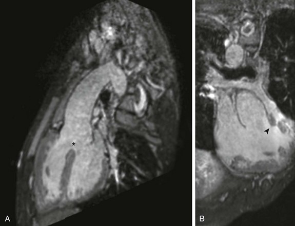
A, Sagittal oblique projection demonstrates the aorta overriding an anterior malalignment ventricular septal defect (asterisk). B, Coronal oblique projection demonstrates the deviation of the conal septum (arrowhead), leading to subvalvar and valvar pulmonary stenosis.
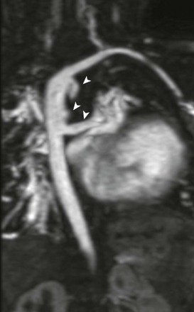
A coronal-oblique maximum intensity projection from a contrast-enhanced magnetic resonance angiogram demonstrates aortopulmonary collaterals (arrowheads). This patient also had a right aortic arch with aberrant left subclavian artery.
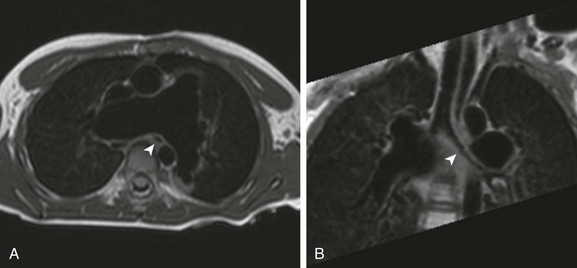
A, An axial image demonstrating markedly dilated branch pulmonary arteries. Note the caliber and position of the left mainstem bronchus (arrowhead) relative to the right pulmonary artery. B, Coronal oblique minimum intensity projection demonstrates a narrowed left mainstem bronchus (arrowhead) as it passes posterior to the proximal right pulmonary artery.
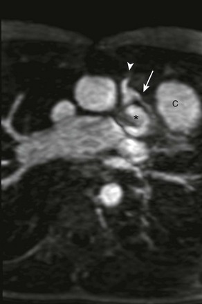
Axial oblique maximum intensity projection from a contrast-enhanced magnetic resonance angiogram shows, with a left anterior descending (LAD) coronary artery (arrow) arising from the right coronary artery (arrowhead) and traveling anterior to the right ventricular outflow tract (asterisk), preventing use of a transannular patch. A right ventricle to pulmonary artery conduit (C) was placed instead, anterior to the LAD coronary artery.
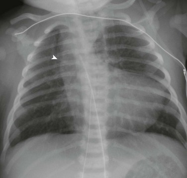
Anteroposterior chest radiograph demonstrates the upturned apex and concavity in the region of the pulmonary artery (a boot-shaped heart). Note the shadow to the right of the trachea (arrowhead), which is indicative of a right aortic arch.
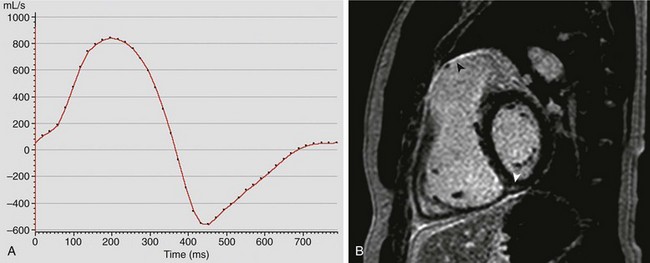
A, Graphical representation (flux vs. time) of antegrade and regurgitant flow in the main pulmonary artery. This patient had severe pulmonary insufficiency, with a regurgitant fraction of 57%. B, Late gadolinium enhancement imaging in the short axis of the ventricles demonstrates enhancement (arrowheads) along the right ventricular outflow tract and at the inferior insertion point of the interventricular septum. See Videos 76-3, 76-4, and 76-5.
Transposition of the Great Arteries
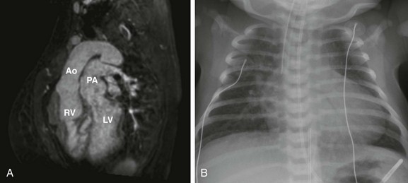
A, Sagittal oblique projection from three-dimensional steady-state free-precession imaging in a 29-year-old woman with {S,D,D} transposition of the great arteries, after an atrial switch procedure, demonstrates parallel outflow tracts, with the aorta (Ao) arising from the right ventricle (RV) and the pulmonary artery (PA) arising from the left ventricle (LV). B, A chest radiograph of a neonate with {S,D,D} TGA prior to repair. As a result of the parallel, anterior-posterior relationship of the great arteries, patients with {S,D,D} transposition of the great arteries have a narrow mediastinum, which, combined with cardiomegaly and increased vascular flow, produces the classic “egg on a string” appearance.
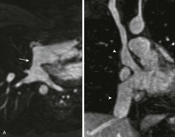
A, Transverse oblique projection demonstrating an unobstructed pulmonary venous baffle (arrow) to the tricuspid valve. B, A coronal oblique projection demonstrating unobstructed pathways from the superior and inferior vena cavae (arrowheads) to the mitral valve. See Video 76-6.
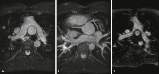
A, Transverse projection demonstrating the pulmonary artery bifurcation anterior to the ascending aorta, with no narrowing of the proximal branch pulmonary arteries. B,Related posts:
![]()
Stay updated, free articles. Join our Telegram channel

Full access? Get Clinical Tree


