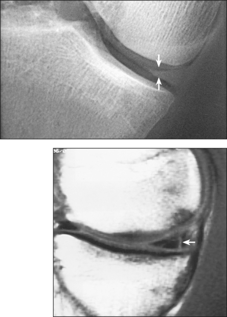
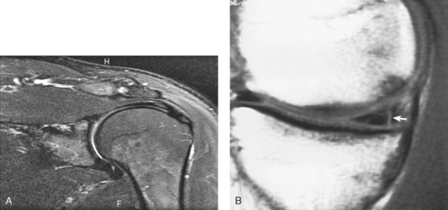
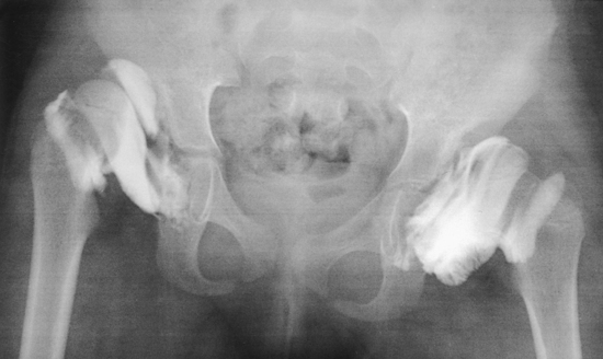
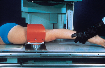
12 The introduction and development of magnetic resonance imaging (MRI) have significantly reduced the number of arthrograms performed in radiology departments. Because MRI is a noninvasive imaging technique, the knee, wrist, hip, shoulder, temporomandibular joint (TMJ), and other joints previously evaluated by contrast arthrography are now studied using MRI (Fig. 12-1). As a result, radiographic contrast arthrography has increasingly specialized functions. A gaseous medium is used in pneumoarthrography, a water-soluble iodinated medium is used in opaque arthrography (Fig. 12-2), and a combination of gaseous and water-soluble iodinated media is used in double-contrast arthrography. Although contrast studies may be made on any encapsulated joint, the shoulder is the most frequent site of investigation. Other joints examined by contrast arthrography include the knee, hip, wrist, and TMJs. • Place the limb in the frame to widen or “open up” the side of the joint space under investigation. This widening, or spreading, of the intrastructural spaces permits better distribution of the contrast material around the meniscus. • After the contrast material is injected, place the limb in the stress device (Fig. 12-3). To delineate the medial side of the joint, place the stress device just above the knee and then laterally stress the lower leg. • When contrast arthrograms are to be made by conventional radiography, turn the patient to the prone position and fluoroscopically localize the centering point for each side of the joint. The mark ensures accurate centering for closely collimated studies of each side of the joint and permits multiple exposures to be made on one IR. The images obtained of each side of the joint usually consist of an AP projection and a 20-degree right and left AP oblique projection. • Obtain the oblique position by leg rotation or central ray angulation (Fig. 12-4). • On completion of these studies, remove the frame and perform lateral and intercondyloid fossa projections. NOTE: Anderson and Maslin1 recommended that tomography be used in knee arthrography. In addition, the technique frequently can be used for other contrast-filled joint capsules.
CONTRAST ARTHROGRAPHY
Overview
SUMMARY OF PATHOLOGY
Condition
Definition
Dislocation
Displacement of a bone from a joint
Joint capsule tear
Rupture of the joint capsule
Ligament tear
Rupture of the ligament
Meniscus tear
Rupture of the meniscus
Rotator cuff tear
Rupture of any muscle of the rotator cuff
Contrast Arthrography of the Knee

Related posts:
Stay updated, free articles. Join our Telegram channel

Full access? Get Clinical Tree


