8 Development of the Abdominal Aorta
K.I. Ringe, S. Meyer
The branches of the abdominal aorta may be divided into three groups: posterior, lateral, and anterior.
8.1 Posterior Branches
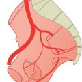
The posterior branches are segmental like the branches of the thoracic aorta.1 The lumbar arteries I–IV remain segmental arteries, but the more important fifth artery forms the main artery for the leg, the common iliac artery. It is comparable to the main vessel supplying the arm, the subclavia, which develops from the sixth cervical segmental artery. Caudal to the fifth lumbar artery, the aorta regresses to the small median sacral artery. The lumbar arteries are not described in detail because their anomalies are comparable to those of the intercostal arteries (Chapter 4). Although the trunk formation of both lumbar arteries from one segment is more common (4%), the origin of two or more lumbar arteries on one side is much more seldom (2%) than in the thoracic region.1

Fig. 8.1 Development of the posterior branches. Schematic.
8.2 Lateral Branches
The lateral branches supply the kidneys and the genital organs. The mesonephric arteries supply the mesonephros, the adrenal glands, and testicles or ovaries. In the second month of pregnancy the adrenal gland is much larger than the kidney and is supplied by three arteries: the superior, middle, and inferior suprarenal arteries. One branch runs from the superior suprarenal artery to the diaphragm and another from the inferior suprarenal artery to the kidney. Later the kidneys grow much faster than the adrenal glands; in the neonate, they are one-third of the kidney weight but in adults only 1/30. Consequently, the suprarenal arteries become less important, and their side branches regress into the main artery. The superior suprarenal artery becomes a small branch of the inferior phrenic artery and the inferior suprarenal artery, a comparably tiny branch of the renal artery. The testicles and ovaries descend, thus elongating their arteries. The origins of these arteries retain their site on the aorta.

Fig. 8.2 Development of the lateral branches. Schematic.
Related posts:


Stay updated, free articles. Join our Telegram channel

Full access? Get Clinical Tree


