33 Development of the Arteries of the Arm
B. Meyer, L. Sonnow
The arteries of the arm show many rearrangements (Fig. 33.1), especially the arteries of the forearm, which develop like the arteries in the leg. The axial artery of the arm is the continuation of the sixth cervical segmental artery (see Chapter 2). This artery has different names: it starts as the subclavian artery, continues as the axillary artery, and is named the brachial artery after passing the posterior axillary fold. In the forearm, the continuation was originally the interosseous artery, which initially supplied the arteries of the fingers. At the next stage, the median artery, which runs with the median nerve, becomes the main artery. In lower mammals, the median artery supplies the finger arteries, and in nonmammals the interosseous artery even remains the main vessel. Only in primates does the final arrangement take place.
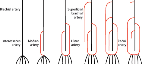
Fig. 33.1 A rough sketch of the development of the arteries of the upper limb. The original system is shown in black; arteries formed at later stages are shown in red.
In human embryos of approximately 18 mm crown–heel length, the ulnar artery as a branch of the brachial artery forms the superficial arterial palmar arch, which supplies the arteries of the fingers. In embryos of 21-mm length, the superficial vessels widen, starting from the axillary region. This superficial brachial artery turns to the lateral side and ends on the dorsal side of the hand. This vessel also connects to the palmar arch and the median artery regresses (embryo of 23-mm length). An anastomosis between the superficial brachial and the brachial artery increases in size, while the proximal part of the superficial brachial artery normally disappears. What remains is the radial artery. This complicated development explains the many variants observed. However, since anastomoses are formed with other additional superficial arteries of the forearm (superficial antebrachial artery), the picture becomes even more confusing. Fig. 33.2 shows the general possibilities, with the different types outlined in Chapter 34 and Chapter 35.1–6
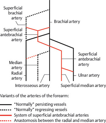
Fig. 33.2 Variants of the arteries of the forearm. Schematic. The solid black line represents “normally” persisting vessels; the dashed black line represents “normally” regressing vessels; the solid red line represents the system of superficial antebrachial arteries; and the dashed red line represents an anastomosis between the radial and median arteries.
Related posts:


Stay updated, free articles. Join our Telegram channel

Full access? Get Clinical Tree


