44 Development of the Arteries of the Head
F. Goetz, A. Giesemann
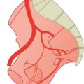
The arteries of the head derive from branchial arteries (I–IV), the early development of which is described in Chapter 1. The cervical segmental arteries (see Fig. 44.1, C1–C6) form an anastomotic chain which later becomes the vertebral artery (see Fig. 44.3; cervical and cerebral vertebral arteries). The corresponding “segmental arteries” of the head (see Fig. 44.1; proatlantal, trigeminal, otic, and hypoglossic artery) are connected with the terminal branch of the internal carotid artery, the cerebral vertebral artery, which bends in a posterior and caudal direction.
The cervical and cerebral vertebral arteries form one continuous artery on each side and the segmental arteries disappear (Fig. 44.2 and Fig. 44.3). Between the trigeminal and hypoglossic artery, the initially paired vertebral arteries merge to form the basilar artery.
At an early stage, a large area of the face is supplied by a branch of the second branchial artery (Fig. 44.2), the stapedial artery. Its name derives from the stapes of the middle ear which forms around this artery. The three main branches of the stapedial artery join the three branches of the trigeminal nerve, and the supraorbital, infraorbital, and mandibular arteries. Later (Fig. 44.3) two anastomoses are formed, one between the maxillary artery from the external carotid artery and the mandibular artery, and the other between the ophthalmic and the supraorbital arteries. The stapedial artery atrophies, and the maxillary artery supplies almost the whole area of the primitive stapedial artery. Only the supraorbital artery remains connected to the ophthalmic artery.
The anomalies shown in Chapter 43 for the maxillary artery can be explained by the embryological development. The original anastomosis between the stapedial and external carotid arteries is medial to the mandibular nerve. As a rule, a second anastomosis is formed lateral to the lateral pterygoid muscle and the first anastomosis disappears.1–12
44.1 Persistent Stapedial Artery (<0.1%)
A persistent stapedial artery is a rare anomaly. This artery originates from the extracranial part of the internal carotid artery, enters the skull medial to the styloid process, runs to the middle ear in an osseous canal, is surrounded by the stapes, and reaches the middle cerebral fossa in front of the canal of the major petrosal nerve and ends as a middle meningeal artery (see also Fig. 44.2).13–19
Cross-sectional imaging findings of a persistent stapedial artery are (1) a linear structure crossing the middle ear over the promontory; (2) an enlarged facial nerve canal or a separate canal parallel to the facial nerve; and (3) absence of the foramen spinosum.20

Fig. 44.1 Early development of the arteries of the head which derive from the branchial arteries. Longitudinal anastomosis of the segmental arteries (C1–C6) forms the vertebral artery. I–IV, branchial arteries.

Fig. 44.2 Persistent trigeminal and hypoglossic arteries. At an early stage, the stapedial artery forms anastomoses to the ophthalmic and middle meningeal arteries.

Fig. 44.3 Later, additional anastomoses (dashed red lines) are formed.
44.2 Anastomoses between Internal Carotid and Basilar Artery
44.2.1 Trigeminal Artery (<0.1%)
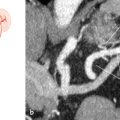
The trigeminal artery branches from the intracranial internal carotid artery within the cavernous sinus, pierces the dorsum sellae frequently and reaches the rostral parts of the basilar artery. It is 2 to 3 cm long (Fig. 44.4). The increased performance of angiographies of cerebral arteries has resulted in the more frequent description of this anomaly.11,21–31

Fig. 44.4 Trigeminal artery (<0.1%). 3D time-of-flight MRA, lateral (a) and anterior (b) MIPs. 1 Trigeminal artery; 2 internal carotid artery; 3 basilar artery.
Related posts:


Stay updated, free articles. Join our Telegram channel

Full access? Get Clinical Tree


