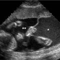
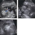
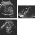
Figure 30.1.1
Normal saline infusion sonohysterogram. A: Sagittal transvaginal view of the uterus demonstrates fluid (*) that was instilled via a transcervical catheter into the uterine cavity. The inner surface of the endometrium (arrowheads), outlined by the fluid, is smooth with uniform thickness. B: The single-layer endometrial thickness (calipers) is slightly more than 1 mm on each side.
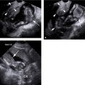
One of the main uses of SIS is in women with postmenopausal bleeding and a thick endometrium on ultrasound. In this setting, endometrial biopsy is generally performed because of suspicion for endometrial cancer, and SIS can help select the best biopsy technique by determining whether the thickening is diffuse (Figure 30.1.2) or focal (Figure 30.1.3). If diffuse, a blind endometrial biopsy is appropriate, whereas hysteroscopic biopsy may be needed if there are focal areas of thickening.
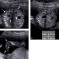
SIS can also prove useful in premenopausal woman. Instillation of saline can identify adhesions (Figure 30.1.4), which appear as linear structures crossing the uterine cavity, or polyps (Figure 30.1.5).
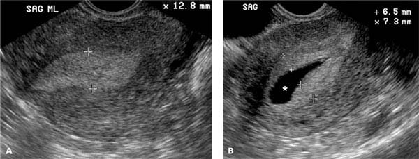
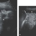
Figure 30.1.2
Saline infusion sonohysterogram demonstrating diffuse endometrial thickening. A: Sagittal transvaginal view of the uterus in a woman with postmenopausal bleeding reveals a thickened endometrium (calipers) measuring 12.8 mm. B: After instillation of saline (*), the endometrium is seen to be diffusely thick (calipers).
Stay updated, free articles. Join our Telegram channel

Full access? Get Clinical Tree


