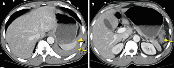
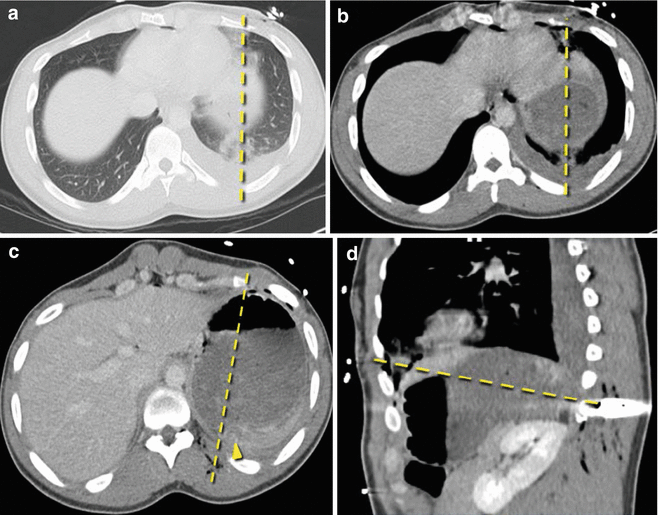
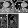
A 62-year-old man who was struck by a car: Computed tomography (CT) images demonstrate the absence of the interposition of lungs between the upper abdominal contents and the chest wall consistent with the dependent viscera sign (arrow). There is intrathoracic herniation of abdominal viscera as demonstrated by visualization of abdominal organs within the pleural space (circle). Additionally, coronal reconstructions demonstrate elevation of left-sided abdominal contents above the left diaphragmatic dome (line)
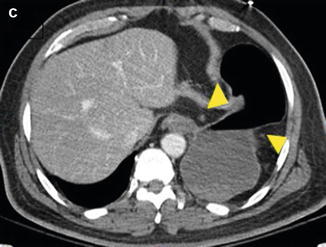
A 62-year-old man who was struck by a car: Computed tomography (CT) image demonstrates the tear in the diaphragm results in a waist-like constriction of the stomach (image C arrowheads). These findings are representative of the collar sign. The patient’s CT findings of a left diaphragmatic rupture were confirmed at surgery
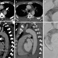
A 25-year-old who suffered a motor vehicle collision: CT images demonstrate a linear band of hypoattenuation through the liver, the band sign (images A and B, dashed line). Once again, we see abdominal organs located within the left pleural space (image A, circle) and the dependent viscera sign (image A, arrow). Abdominal contents are located above the level of the diaphragm (image B, arrowheads). The patient’s CT findings of bilateral diaphragmatic rupture were confirmed at surgery. Bilateral diaphragmatic injury can occur in 5–8 % of cases of blunt trauma with diaphragmatic injury. Consequently, a careful inspection of the contralateral hemidiaphragm should be pursued once a diaphragmatic injury is found
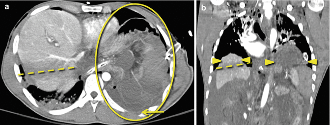
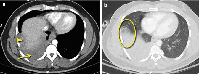
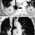
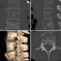
A 33-year-old in motor vehicle collision: Axial CT images demonstrate the dangling diaphragm sign as evidenced by the inward curling of the diaphragm from its normal course (images A and B, arrows). Additionally, there is thickening of the injured diaphragm (images A and B, arrows). The collar sign is also present (images A and B, arrowheads)
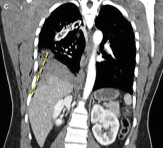
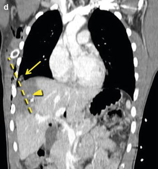
A 33-year-old in motor vehicle collision: The topogram demonstrates apparent elevation of the left hemidiaphragm (image C, dashed line). The coronal CT reformations confirm elevated abdominal organs within the left thorax ≥4 cm above the level of the level of the right diaphragmatic dome (image D, dashed line). Diaphragmatic rupture was confirmed at surgery
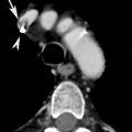
A 47-year-old man stabbed in the left flank: Axial CT images demonstrate contiguous injuries above and below the diaphragm which raise the suspicion of diaphragmatic injury. There is a left hemothorax (image A, arrow), hemoretroperitoneum with blood tracking along the gastrosplenic ligament (image B, arrow), and a focus of free gas abutting the spleen (image A, arrowhead)
A 34-year-old man with gunshot wound to the right chest: Axial CT images demonstrate a pulmonary contusion (image B, circle), hemothorax (image A, arrow), and hepatic laceration (image A, arrowhead). Contiguous injury on both sides of the diaphragm suggests diaphragmatic injury
A 34-year-old man with gunshot wound to the right chest: Multiplanar reconstruction demonstrates the wound tract (image C, dashed line) traversing the right hemidiaphragm with injuries on both sides of the diaphragm. This case illustrates the value of coronal images to find the wound tract. The patient’s diaphragmatic injury was confirmed at surgery

A 21-year-old man with gunshot wound to the right chest: Axial CT image shows a liver laceration with active extravasation of contrast (image A, arrow) and an associated subcapsular hematoma. Axial lung windows demonstrate a pulmonary contusion (image A, arrowhead) and trace pneumothorax. Illustrative wound tracts of metallic shot pellets traverse the right hemidiaphragm. No diaphragmatic discontinuities are seen on CT, but these findings are consistent with diaphragm laceration
A 21-year-old man with gunshot wound to the right chest: Once again we see the benefit of multiplanar reformations. Coronal images show that the wound tract (dashed line) courses through the right hemidiaphragm. Contiguous injury above and below the right hemidiaphragm was also demonstrated as lung injury from the bullet tract can be seen above the diaphragm (arrow) and active extravasation within the liver is seen along the path of the shot (arrowhead). Right diaphragmatic injury was confirmed at surgery
A 28-year-old man with a gunshot wound to the left upper quadrant: Axial images demonstrate pulmonary contusion. The bullet tract (images A and B, dashed line) traverses the left hemithorax and left upper quadrant. The wound tract (image C, dashed line) also courses inferiorly through the abdomen, and there is evidence of hemoperitoneum (arrowhead). Contiguous injuries above and below the diaphragm are concerning for diaphragmatic injury. Sagittal reformations show the wound tract (image D, dashed line) coursing from above the diaphragm into the abdomen. Left diaphragmatic injury was confirmed at surgery
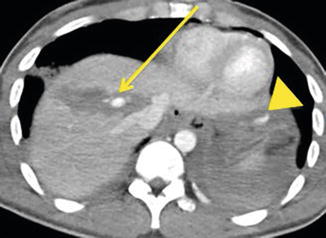
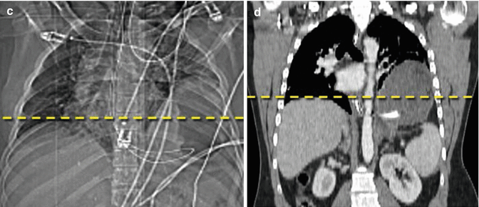
GSW with tract through the upper abdomen; image shows active extravasation from hepatic laceration (arrow) and below the left hemidiaphragm (arrowhead)
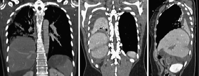
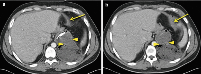
MVC with large right diaphragmatic defect, herniation of the liver into the left thorax, band, and collar signs
References
Barbiera F, Nicastro N, Finazzo M, Lo Casto A, Runza G, Bartolotta TV, Midiri M (2003) The role of MRI in traumatic rupture of the diaphragm. Our experience in three cases and review of the literature. Radiol Med 105(3):188–194PubMed
Bergin D, Ennis R, Keogh C, Fenlon HM, Murray JG (2001) The “dependent viscera” sign in CT diagnosis of blunt traumatic diaphragmatic rupture. AJR Am J Roentgenol 177(5):1137–1140CrossRefPubMed
Related posts:
Stay updated, free articles. Join our Telegram channel

Full access? Get Clinical Tree


