Heller myotomy (partial thickness incision of lower esophageal sphincter)
– Partial (Toupet) fundoplication often incorporated into myotomy procedure
Per-oral endoscopic myotomy (POEM) procedure
– Relief of symptoms with fewer complications
(Left) Upright frontal esophagram shows a dilated esophagus with an abrupt taper (“bird beak”) just above the gastroesophageal (GE) junction . Note the absent gastric air bubble and the fluid-barium level within the esophagus.
(Right) Esophagram shows a grossly dilated, tortuous esophagus with a “sigmoid” appearance. This is an example of longstanding achalasia.
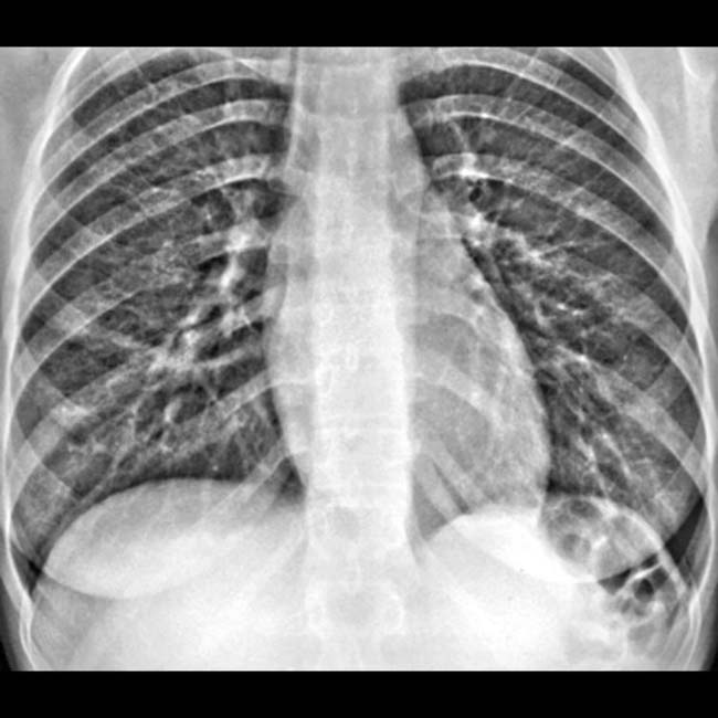
(Left) Upright chest radiograph shows an absent air-fluid level in the stomach of a 28-year-old woman with a recent onset of dysphagia and halitosis.
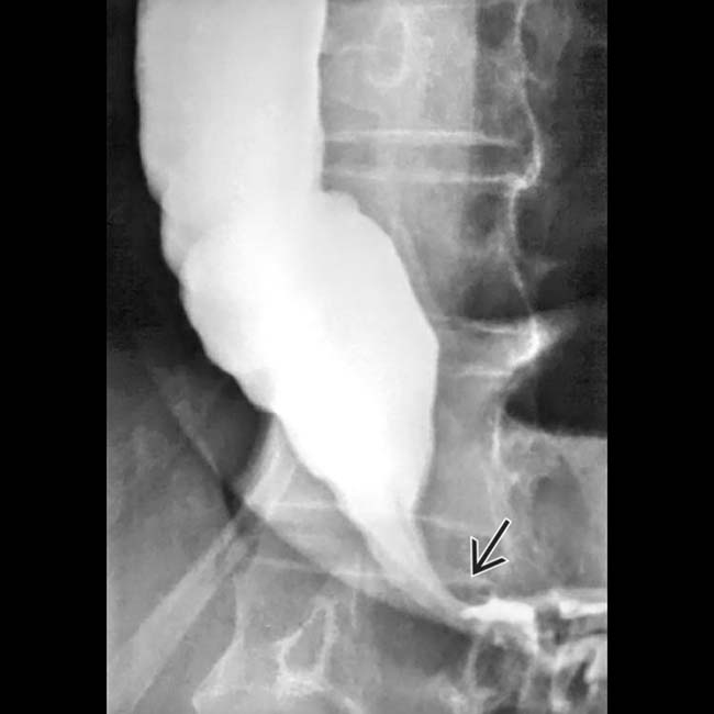
(Right) Esophagram in the same young woman shows marked dilation of the esophageal lumen ending in a smoothly tapered “bird beak” deformity .
TERMINOLOGY
Synonyms
• Cardiospasm
Definitions
• Primary esophageal motility disorder due to defective neural stimulation of lower esophageal sphincter
IMAGING
General Features
• Best diagnostic clue
“Bird beak” deformity: Dilated esophagus with smooth, symmetric, tapered narrowing at esophagogastric region
• Morphology
Grossly dilated esophagus with smooth tapering at lower end of esophagus
• Other general features
Classified based on etiology
– Primary (idiopathic)
– Secondary (pseudoachalasia)
Manometric characteristics of achalasia
– Absence of primary peristalsis
– Increased or normal resting lower esophageal sphincter (LES) pressures
– Incomplete or absent LES relaxation on swallowing
Variants of achalasia: Atypical manometric findings
– Early: Characterized by aperistalsis with normal LES pressure
– Vigorous: Simultaneous high amplitude and repetitive contractions
– Both variants are transitional and finally evolve into classic achalasia
 Transient flow of fluid into stomach when hydrostatic pressure of fluid column exceeds tonic LES pressure


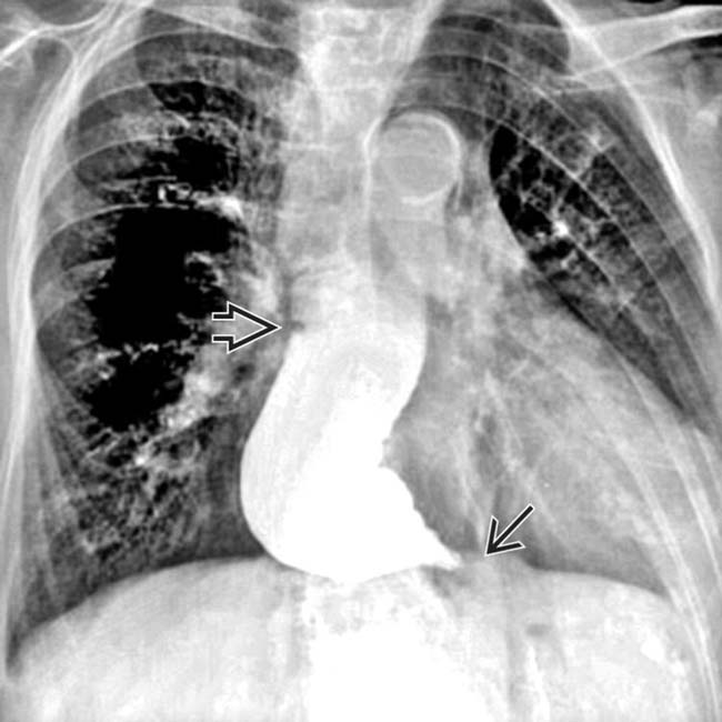
 . Note the absent gastric air bubble and the fluid-barium level
  within the esophagus.
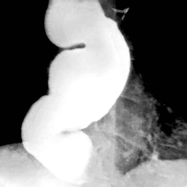
 .
.


 Primary achalasia
 Secondary achalasia (pseudoachalasia)


