Confirmed by contrast esophagram or CT, which are complementary
• Cervical esophageal perforation (EP)
Subcutaneous or interstitial emphysema; neck and mediastinum
Consider perforation of Zenker diverticulum
• Thoracic EP
Chest film: Pneumomediastinum, pleural effusion
• EP of intraabdominal segment of distal esophagus
Abdominal plain film: Pneumoperitoneum
• EP near GE junction
Extravasated contrast from left lateral aspect of distal esophagus into mediastinum, sometimes pleural space, and rarely abdomen (never abdomen alone)
• CT shows extraesophageal air in almost all cases, fluid and contrast medium in most
• Intramural EP: Extravasated gas and contrast remain within esophageal wall
Much better prognosis
• Esophagography: Technique
Esophagram: Videofluoroscopic and rapid sequence filming
Nonionic water-soluble contrast media (e.g., Omnipaque) initially, followed with barium if no leak or fistula seen
Barium (or CT) may detect small leak not visible initially
TOP DIFFERENTIAL DIAGNOSES
• Esophageal diverticulum
• Esophageal ulceration
• Boerhaave syndrome
• Postoperative state, esophagus
• Tracheobronchial aspiration
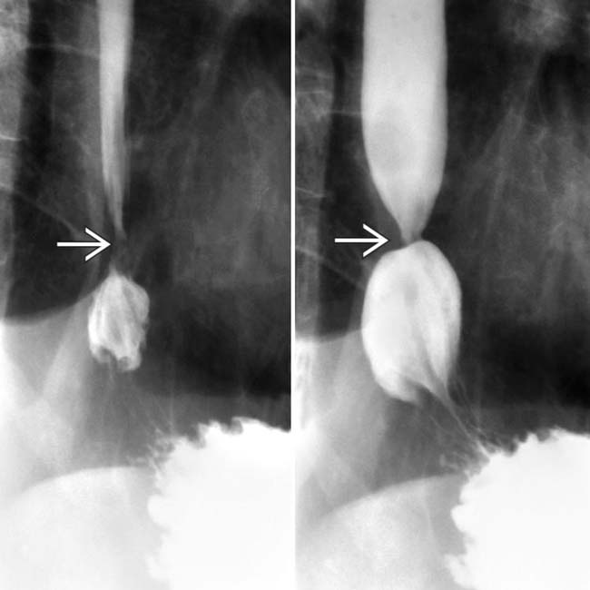
(Left) Barium esophagrams reveal a tight stricture at the gastroesophageal (GE) junction . Due to concern for Barrett metaplasia or early cancer, an endoscopic biopsy of the lesion was performed following balloon dilation of the stricture.
(Right) Postbiopsy esophagram in the same patient illustrates a focal intramural barium collection , indicating a localized perforation. These intramural perforations will usually heal spontaneously.
(Left) Esophagram in a 62-year-old man with a history of laparoscopic hiatal hernia repair, now presenting with subsequent chest pain and fever, demonstrates mediastinal and abdominal extraluminal collections of gas and contrast material.
(Right) Axial CECT in the same patient reveals mediastinal and abdominal extraluminal collections of gas and contrast material, indicating perforation near the GE junction. Surgical drainage was successful.
TERMINOLOGY
Abbreviations
• Esophageal perforation (EP)
Synonyms
• Esophageal rupture or transection
Definitions
• Transmural esophageal tear
IMAGING
General Features
• Best diagnostic clue
Diagnosis depends on high degree of suspicion and recognition of clinical features
– Confirmed by contrast esophagram or CT
• Location
Cervical EP: Posterior wall of esophagus at level of cricopharyngeus muscle
– Or through Zenker diverticulum
Thoracic EP: At or near gastroesophageal (GE) junction
– Areas of anatomic narrowing, sites of extrinsic compression by aortic arch or L main bronchus
At or above benign or malignant strictures
Site of ruptured anastomosis or after esophageal surgery
Radiographic Findings
• Radiography
Cervical EP: Anteroposterior, lateral films of neck


 Nonionic water-soluble contrast media (e.g., Omnipaque) initially, followed with barium if no leak or fistula seen

 . Due to concern for Barrett metaplasia or early cancer, an endoscopic biopsy of the lesion was performed following balloon dilation of the stricture.
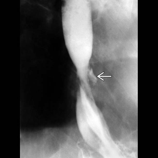
 , indicating a localized perforation. These intramural perforations will usually heal spontaneously.
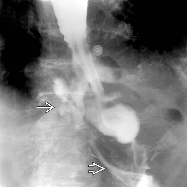
 and abdominal
  extraluminal collections of gas and contrast material.
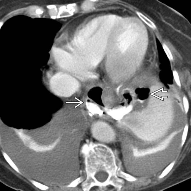
 and abdominal
  extraluminal collections of gas and contrast material, indicating perforation near the GE junction. Surgical drainage was successful.


 Extravasated contrast from left lateral aspect of distal esophagus into mediastinum, sometimes pleural space, and rarely abdomen (never abdomen alone)


