4 Evaluation of Flexor Tendon Lacerations
♦ Setup
• The patient is seated with the hand placed palm upward on the examination table.
• The patient is usually holding the injured finger extended because of the nature of the injury. If this is not the case, the finger is manually extended.
• Typically the smallest probe is best for finger examination. Although it provides the smallest field of view, it gives the highest resolution. It is typically sufficient even into the forearm when looking at structures as small as a tendon.
♦ Landmarks
• The flexion creases of the fingers are excellent landmarks.
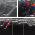
• The flexor tendons can be identified more easily when the patient flexes and extends the fingers. The flexor digitorum superficialis (FDS) splits into two slips at approximately the level of the metacarpophalangeal (MCP) flexion crease.
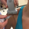
♦ Probe Positioning
Axial
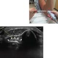
• The probe should be 90 degrees to the long axis of the finger (Fig. 4.1). The probe must be maintained tangentially perpendicular to the surface of the finger.
• The center arrow of the probe should be just over the center of the digit to properly image the flexor tendons.
• It is typically easier to identify both the FDS and the flexor digitorum profundus (FDP) tendons in the axial plane proximal to the MCP flexion crease, around the level of the A1 pulley.

Related posts:
Stay updated, free articles. Join our Telegram channel

Full access? Get Clinical Tree


