9 Evaluation of Lateral Epicondylitis
♦ Setup
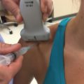
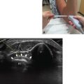
• The patient should be seated, facing the operator, with the elbow resting on the table (Fig. 9.1).
• The elbow should be flexed to 90 degrees, with the forearm pronated. Local anesthesia can be used to minimize pain.
• Either a standard shoulder probe or a narrower probe should be used. A larger probe gives a wider field of view but requires a flatter injection angle.

Fig. 9.1 Elbow flexed 90 degrees, forearm pronated.
♦ Landmarks
The following landmarks should be noted (Fig. 9.2):
• Lateral epicondyle
• Radial head: supinating and pronating the forearm will confirm the location
• Extensor carpi radialis brevis (ECRB) insertion: just proximal and anterior to the lateral epicondyle

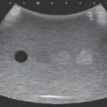
Fig. 9.2 The lateral epicondyle (LE) and radial head (RH) should be palpated. ECRB, Extensor carpi radialis brevis.
♦ Probe Positioning
Axial
• The probe should be positioned 90 degrees to the long axis of the forearm.
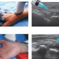
• The probe should be centered over the lateral epicondyle and moved just anterior (Fig. 9.3).
• The probe should be perpendicular to the floor.

