Fig. 14.1
HyCoSy with water and air: this image demonstrates air echogenic areas with a background of echo-free areas. The air bubbles are seen to move through the tube to demonstrate patency
In the mid-1980s, an ultrasound contrast agent named Echovist® was being trialled for use in echocardiography. Due to its echogenic properties, Echovist® revolutionised the visualisation of the fallopian tubes using HyCoSy. Echovist® consists of galactose particles suspended in an aqueous galactase solution. Echovist is no longer available, and SonoVue®, a second-generation agent, is now commonly used. The SonoVue® kit consists of a lyophilised powder which is mixed vigorously with normal saline to form the injectable contrast media. SonoVue consists of microbubbles of sulphur hexafluoride. The interface between the sulphur hexafluoride bubble and aqueous medium acts as a reflector of the ultrasound beam, thus enhancing blood echogenicity and increasing contrast between the blood and the surrounding tissues (Fig. 14.2).
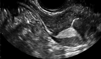
Fig. 14.2
HyCoSy with SonoVue® dye showing dye in the uterine cavity and the right tube
The contrast agent produces a hyperechoic appearance on transvaginal ultrasonography. The contrast media are detected first in the uterine cavity, proximal and then distal fallopian tubes (if they are patent). Tubal patency is demonstrated by visualising intratubal flow for 5–10 s using B-mode scanning and until peritoneal spill is detected around the ovaries [14].
Below we outline a suggested technique for performing the procedure. There are variations to this technique, as well as inclusion and exclusion of steps that may not be routinely performed by other operators.
As HyCoSy is often performed as an outpatient procedure, it is imperative that clinicians performing this procedure remember the basics of good bedside manner, effective communication and making the patient feel at ease. Most patients will be apprehensive about the possible findings but also the anticipated discomfort. Operators performing HyCoSy should be proficient in transvaginal ultrasonography and placement of transcervical catheters and possess the relevant clinical experience and skills to perform this investigation.
It is good practice to issue patients with an information leaflet (some time before the procedure) outlining the procedure so that they have some idea of what to expect when they attend. Leaflets can also inform patients of what to do pre-procedure and expect post-procedure and whom to contact in the event of any complications.
Some operators will perform a urinary beta-HCG test to exclude pregnancy prior to commencing the procedure, although as HyCoSy is performed in the follicular phase of the cycle, this isn’t done routinely.
The Technique
1.
After gaining verbal consent and a brief description of the procedure, the patient is placed into the dorsal lithotomy position.
2.
A warmed, sterile and well-lubricated Cusco’s (bivalve) speculum (of the appropriate size for the patient) is then carefully and slowly inserted into the vagina in order to visualise the cervix. Occasionally, the cervix may not be easily identified, and gently changing the angle of direction of the speculum may help with this.
3.
Once the cervix is identified, it is cleaned with an aseptic solution.
4.
The authors recommend the use of a flexible balloon catheter and not the previously used metal cannulae. Foleys catheters have also been employed at this stage. The insertion of the catheter does not routinely require the use of a tenaculum; however, if tenaculum use is required, then the authors suggest a paracervical block with 1 % lignocaine prior to grasping the cervix or only blocking the anterior lip when the tenaculum is applied.
5.
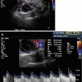
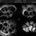
If a balloon catheter is used, then the authors recommend intracervical, as opposed to intrauterine, balloon dilatation. It has been demonstrated that this causes less pain, and less contrast media are required in this way too [15]. The balloon can be inflated with air or sterile water. This also allows visualisation of the lower end of the uterine catheter. If the catheter is found to be placed in the uterine cavity under ultrasound guidance, this can be withdrawn into the cervical canal. Figure 14.3 shows the balloon in the uterine cavity – this is occasionally done if the catheter does not appear to be well fixated in the cervical canal (and therefore prevents it from falling out).
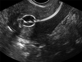
Fig. 14.3
HyCoSy catheter in cavity – ideally the catheter should be in the cervical canal. Occasionally, it is placed in the cavity to prevent displacement during the procedure
6.
Once the catheter is in situ and secure, the speculum (and tenaculum if applied) can be gently removed, ensuring the catheter is not dislodged. The patient is then forewarned that the transvaginal ultrasound probe will be inserted.
7.
At this stage, the authors perform a conventional B-mode transvaginal scan to assess the uterus, ovaries and pouch of Douglas. The correct placement of the catheter balloon can also be checked at this point. Alternatively, a conventional scan can be performed after step 1(before the catheter is introduced).
8.
After warning the patient, the contrast medium can be injected slowly and steadily. It is important to remember that the uterus is pressure sensitive, and as such, excessive rates and/or volumes of injecting will result in unnecessary patient discomfort. Beware that blocked fallopian tubes may increase the pain experienced by the patient. The authors suggest using no more than 10 ml of contrast media. If the balloon has been inflated correctly, there should be no leakage, and evaluation of the uterus and both tubes should be possible using less than 10 ml. In the author’s experience, 2–5 ml is sufficient for demonstrating tubal patency.
9.
Tubal patency is assessed by demonstrating flow along the entire length of the tube or by streaming at the cornual end for at least 10 s with spill into the pouch of Douglas [16].
10.
A detailed examination of the uterus is performed by scanning slowly and systematically from the cervix to fundus. Any relevant lesions (e.g. submucous leiomyoma) can be closely analysed and relevant images produced.
11.
Each tube is followed, in turn, until spill is visualised adjacent to the ovary.
12.
Strict criteria must be adhered to in order to ensure that the fallopian tube is followed in its entirety, before it is considered to be patent. Any delay in tubal fill and/or spill must be appropriately documented. Any apparent distortion of the tubal diameter or tubal course must also be documented and preferable supplemented with the use of images/videography.
13.
This could be followed by assessment of the uterine cavity with normal saline to exclude endometrial polyp or submucous fibroids.
HyCoSy (and HSG) has the significant advantage over laparoscopy of being outpatient-based (office) investigations without a need for general anaesthesia. There is no risk of visceral or vascular injuries. Patients do not need to be fasted for either procedure, and both the patient and her partner can be present whilst the investigation is being performed.
Unlike HSG, HyCoSy does not involve the use of ionising radiation and iodine-based contrast media or the use of radiology services – it can be performed by a gynaecologist/specialist in reproductive medicine, obviating the need for a radiologist. As an ultrasound-based investigation, other pelvic structures can be assessed simultaneously. HSG may preclude the need for laparoscopy in some cases, thereby improving patient satisfaction and preventing the need for invasive investigations.
Related posts:
 Ultrasound in Reproductive Medicine: Is It Safe?
 Ultrasound and PCOS
 Focused Ultrasound for Treatment of Fibroids
 Ovarian Reserve and Ovarian Cysts
 Virtual Hysterosalpingography: A New Diagnostic Technique for the Study of the Female Reproductive Tract
 The Normal Ovary (Changes in the Menstrual Cycle)
Stay updated, free articles. Join our Telegram channel

Full access? Get Clinical Tree


