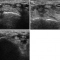
Fig. 2.1
First dorsal wrist compartment. The first osseofibrous tunnel is located on the radial side of the wrist (a). It contains two tendons, the abductor pollicis longus (Abd l) and the extensor pollicis brevis (Est b), both of which originate deep in the ulnar side of the forearm, from the dorsal aspects (middle third) of the ulna, radius, and interosseous membrane (b). Proceeding toward the periphery (c, d) they become superficial and cross over the extensor carpi radialis longus (Est rlc) and brevis (Est rbc)—a point known as the proximal intersection (e, f)—before entering the first osseofibrous tunnel
As they extend peripherally, they become more superficial (Fig. 2.1c, d), and their tendons cross over those of the long and short radial extensors of the wrist—a point referred to as the proximal intersection (Fig. 2.1e, f)—before entering the first osseofibrous tunnel. At the level of the styloid process of the radius, the first compartment tendons are stabilized by a retinaculum, which is depicted sonographically as a hypoechoic band encircling the tendons and attached to the bone (Fig. 2.2). The abductor pollicis longus inserts into the lateral base of the first metacarpal and the extensor pollicis brevis into the dorsal base of the proximal phalanx of the thumb.
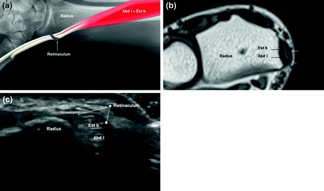
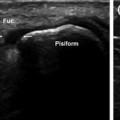
Fig. 2.2
Retinaculum of the first compartment: a Schematic diagram; b Axial T1-weighted MRI scan. At the level of the styloid process of the radius, the abductor longus (Abd l) and extensor brevis (Est b) of the thumb are stabilized by a retinaculum (a, b) (white arrows), which is visualized on sonography (c) as a hypoechoic structure encircling the two tendons that is attached to the bone
The first compartment is characterized by frequent anatomic variations [6]. For example, the abductor longus tendon often has multiple terminal laminae instead of a single insertion (Fig. 2.3). Another clinically significant variant is the presence of an additional septum that divides the first tunnel longitudinally into two parts. In this case, pathological changes may be confined to only one of the two tendons in the compartment (generally the extensor pollicis brevis) (Fig. 2.4) (Table 2.1).
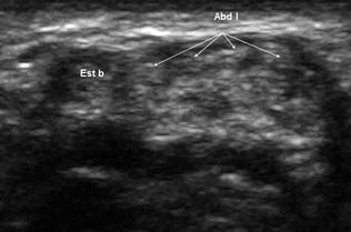
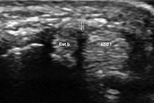
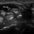
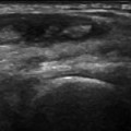
Fig. 2.3
First dorsal wrist compartment, anatomic variant. The insertion of the abductor longus is multilamellar rather than single
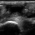
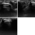
Fig. 2.4
First dorsal wrist compartment, anatomic variant. The first osseofibrous tunnel is divided into two parts by a vertical fibrous septum (arrow)
Table 2.1
Insertions and actions of the extensor tendons
Insertion | Function | |
|---|---|---|
Abductor pollicis longus | First metacarpal, base | Moves the first metacarpal dorsally and radially; helps extend and abduct the wrist |
Extensor pollicis brevis | Proximal phalanx of the thumb, dorsal part | Extends the proximal phalanx; helps extend and abduct the wrist |
Extensor carpi radialis longus | Second metacarpal, dorsal part of the base | Extends the wrist, abducts the hand |
Extensor carpi radialis brevis | Third metacarpal, dorsal part of the base; some fibers insert on bases of the second and fourth metacarpal | Extends the wrist, abducts the hand |
Extensor pollicis longus | Distal phalanx of the thumb | Extends the distal phalanx; helps extend and abduct the wrist |
Extensor digitorum communis | Middle and distal phalanges (bases) of the index, middle, ring, and little fingers | Extends the index, middle, ring, and little fingers; helps extend the wrist |
Extensor indicis proprius | Middle phalanx (ulnar side) of the index finger | Extends the proximal phalanx of the index finger; helps extend the wrist |
Extensor digiti minimi proprius |