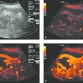
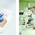
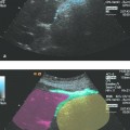
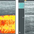
Extravascular Use of Ultrasound Contrast Agents Ultrasound contrast agents (UCAs) are widely used in everyday clinical practice. The majority of indications are for their intravenous use. As early as 1986, however, the first study was published on the extravascular or intracavitary use of UCAs in the renal collecting system.1 The first in vivo reports were published in 2000.2 Since then, studies have been published on the following applications, which fall under the general heading of extravascular contrast-enhanced ultrasound (EVCEUS): Voiding sonography (excretory sonography) Hysterosalpingo-contrast sonography (HyCoSy) Biliary tract: via endoscopic retrograde cholangiography (ERC)3 via percutaneous transhepatic cholangiography and drainage (PTCD)4 Fistulography5 Analogously to the use of conventional radiographic contrast media, UCAs can also be used to improve the visualization of physiologic cavities (e.g., the pleura and peritoneal cavity) and nonphysiologic cavities (e.g., abscesses and cystic lesions), especially during interventional procedures. Imaging of the upper and lower gastrointestinal tract can also be enhanced with UCAs administered by swallowing, injection, or enema. UCAs are approved for intravascular use. They have not been approved for any other indications except voiding sonography with SHU508A (Levovist, Bayer Schering Pharma). Thus, the benefit to the patient must be weighed against possible risks before these agents are used for extravascular imaging, and detailed informed consent must be obtained. To date only a few complications have resulted from the intravenous use of UCAs. In a retrospective multicenter Italian study that included more than 23,000 examinations, the absolute number of adverse events was 29 (0.0086%), 2 of which were severe (1 bronchospasm, 1 allergic shock). No fatal events occurred.6 Thus, given the very low complication rate of intravenous use and the very low dose used in extravascular applications, it is reasonable to conclude that complications are extremely rare during the extravascular use of UCAs. Intravenous UCAs are administered in an extremely low dose. For example, 1 mL of SonoVue (Bracco Imaging) contains 1 to 5 × 10⁸ microbubbles, and 1.2 mL is adequate for intravenous imaging on modern ultrasound machines. UCAs are generally diluted for extravascular use. Since this is not an approved application, no official dose recommendations are available. When a contrast agent is injected at a local concentration that is too high, it produces a shadowing artifact that may obscure image details distal to the ultrasound transducer.7 The administered dose must be reduced, therefore. If too much contrast agent has already been injected, higher-energy ultrasound pulses can be transmitted to reduce the concentration of the microbubbles to a reasonable level. In the few studies on voiding sonography (vesicoureteral reflux) that have been published to date, concentrations of 1 mL SonoVue in 120 to 210 mL of saline solution (calculated for 2- to 5-year-old children) were injected into the bladder of the pediatric patients.8,9 This represents a dilution factor of 100 to 200. For the oral administration of UCAs, 2 to 4 drops of SonoVue are mixed with 200 mL of tap water. For esophageal enhancement the patient can take one mouthful while sitting up, then lie back and swallow the solution while supine. For gastric enhancement all of the solution is swallowed (personal empirical recommendations). For lower distribution volumes (e.g., in the biliary tract), a reasonable dilution is 0.1 mL of SonoVue per 20 mL of saline solution.4 Very large distribution volumes (ascites, large pleural effusions) require a higher contrast dose, and one whole ampule of SonoVue should be injected. If the main goal of the investigation is to determine whether or not contrast medium can enter a cavity (fistulography, vesicoureteral reflux), higher concentrations may be used. The main concern in this case is not image aesthetics but the appearance (or nonappearance) of the contrast medium. In this chapter we will present the most important extravascular applications of UCAs while referring the reader to the appropriate chapters. Vesicoureteral reflux is a relatively common disorder in children and may lead to the deterioration of renal function, so that early diagnosis is essential. Voiding sonography determines whether intravesical contrast medium enters the pyelocalyceal system during micturition, which would confirm vesicoureteral reflux. This requires catheterization of the urethra, generally followed by the injection of a contrast agent. Levovist has been approved for this indication. SonoVue offers many technical advantages over Levovist but has not been approved. Voiding sonography had advantages over radiography in its ability to define renal anatomy without exposure to ionizing radiation. It should also be noted that vesicoureteral reflux is not a constant phenomenon and may elude detection by a radiographic “snapshot.” The male urethra can be imaged by perineal ultrasound to detect urethral stenosis.10 Female infertility tests may include the assessment of tubal patency. Laparoscopy with dye insufflation is considered the gold standard but is invasive. Another method is radiographic hysterosalpingography. Hysterosalpingo-contrast sonography (HyCoSy) was introduced at a relatively early stage to avoid radiation exposure. It did not employ commercial contrast medium but involved the intrauterine injection of saline solution that was shaken to create air bubbles. But several more recent studies have shown that the use of UCAs can improve the test and increase its sensitivity.11–13 Antibiotic prophylaxis is given before the examination (e.g., 200 mg doxycycline 30 minutes before the test or 500 mg azithromycin daily 2 days before the test continued until the day of the test). An 8F balloon catheter is introduced into the cervix. Then SonoVue is diluted with saline solution (1.2 mL SonoVue in 5–10 mL saline) and injected into the uterine cavity.14 Paracentesis is necessary in patients who have diuretic resistance or experience adverse effects from diuretics (e.g., exacerbation of preexisting renal failure). When an ultrasound contrast agent is injected into the peritoneal cavity, the agent is distributed throughout all communicating spaces. This effect is supported by patient movements (e.g., walking around). If septa are present, they will confine the agent to localized areas. A whole ampule should be injected due to the high distribution volume, and the agent does not have to be diluted. Foschi et al reported on seven patients with cirrhotic ascites and pleural effusion. In five patients they were able to document the passage of UCA from the peritoneal to the pleural cavity. This allowed them to confirm the presumptive diagnosis of hydrothorax, which was reconfirmed by nuclear medicine scans. These findings made it easier or possible to proceed with operative treatment. The authors administered two ampules of SonoVue.15 Three-dimensional imaging methods can be used (▶ Fig. 31.1).
31.1 Approved Indications
31.2 Contraindications and Complications
31.3 Technique
31.4 Use of Ultrasound Contrast Agents in Physiologic Body Cavities
31.4.1 Voiding Sonography for the Detection of Vesicoureteral Reflux
31.4.2 Contrast-Enhanced Ultrasound for Evaluating Tubal Patency
31.4.3 Imaging the Peritoneal Cavity with UCAs (for Detection of Ascites)
Stay updated, free articles. Join our Telegram channel

Full access? Get Clinical Tree


