26.1 Normal: OM 30° view
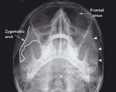
The ‘elephant’s trunk’ of the zygomatic arch (white line and arrowheads) is clearly seen on this view. Note also the frontal air sinuses and the odontoid peg
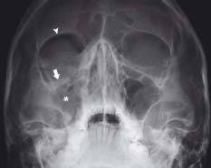
The thin orbital floor (arrow) is depressed with opacification of the maxillary sinus (*) due to blood. Air entering the orbit from the maxillary sinus gives rise to the ‘black eyebrow’ sign (arrowhead)
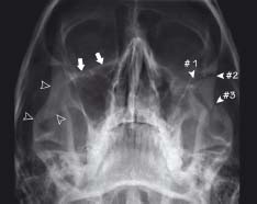
There is a complex fracture involving the orbital floor (#1), lateral orbital wall (#2) and zygomatic arch (#3). Note the normal orbital floor (arrows) and the normal zygomatic arch (open arrowheads) on the right
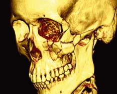
This CT of the same patient as in fig 26.3 reveals a more complex fracture than is appreciated on plain XR. CT can be a useful planning tool before facial surgery
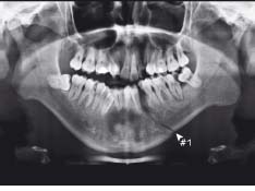
Blunt trauma to this patient’s jaw has caused an obvious fracture (#1). A second fracture should be suspected and further views may be required
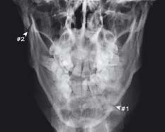
On this view of the same patient as in fig 26.5, the first fracture (#1) is less obvious, however a second fracture (#2) is clearly seen which in hindsight is visible on the OPG
Face anatomy seen on XR
There are several standard plain XR views used to demonstrate the bony anatomy of the face. Structures of the face are anatomically complex, and CT may therefore be required for a more complete assessment of facial fractures or other pathology.
- Occipitomental view (OM) –
Related posts:

Stay updated, free articles. Join our Telegram channel


Full access? Get Clinical Tree


