6 Factors that influence the Doppler spectrum
FACTORS THAT INFLUENCE THE DOPPLER SPECTRUM
Blood flow profile
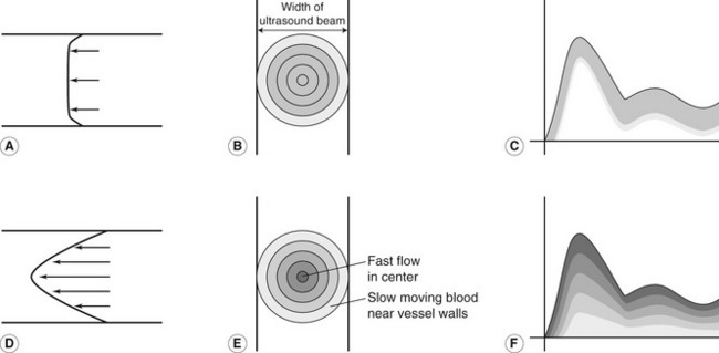
The Doppler spectrum displays the frequency content of the signal along the vertical axis, with the relative brightness of the display relating to the proportion of back-scattered power at each frequency, and the time along the horizontal axis. The velocity profiles seen within arteries can be quite complex and will vary over time, as discussed in Chapter 5. The frequency content displayed in the Doppler spectrum will depend on the velocities of the cells present within the blood. If we assume that the vessel is uniformly insonated by the Doppler beam, all the different velocities of blood present within the vessel will be detected and displayed on the spectrum. If blood is traveling with a blunt flow profile, most of the blood cells will be moving with the same velocity, and the spectrum will show only a small range of frequencies (Fig. 6.1A–C). If, however, the blood is traveling with a parabolic flow profile, then the blood in the center of the vessel will be traveling faster than that near the vessel walls and therefore the Doppler spectrum will display a wide range of frequencies (Fig. 6.1D–F).
The spread of frequencies present within the spectrum at a given point in time is known as the degree of spectral broadening. Figure 6.1 shows the way in which the degree of spectral broadening depends on the velocity profile of the flow being interrogated, with greater spectral broadening seen in Figure 6.1F than in Figure 6.1C. The presence of turbulent flow (e.g., as a result of a stenosis) will increase spectral broadening, as the blood cells will be traveling with different velocities in random directions (see Fig. 5.21). Therefore, increased spectral broadening may indicate the presence of disease. However, the degree of spectral broadening can also be influenced by Doppler instrumentation, and this is known as intrinsic spectral broadening (ISB: discussed later in this chapter).
Nonuniform insonation of the vessel
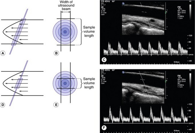
The examples of idealized spectra given in Figure 6.1 assume that the beam evenly insonates the whole cross-section of the blood vessel in order to detect the correct proportions of all the blood velocities present. This is, however, an unrealistic situation as the Doppler beam can be quite narrow (of the order of 1–2 mm wide) and therefore may insonate only part of the artery or vein. If the beam passes through the center of the vessel (Fig. 6.2A), only part of the flow near the vessel walls (i.e., near the anterior and posterior walls) will be detected. The blood flow along the lateral walls will not be detected as it is not insonated by the Doppler beam. Therefore, in the presence of parabolic flow, the low-velocity flow near the walls will only be partially detected and the Doppler spectrum will no longer truly represent the low-velocity flow present within the vessel.
Sample volume size
The size and position of the sample volume, which can be controlled by the operator, will also affect the proportion of the vessel insonated. A small sample volume placed in the center of a large vessel may not detect any of the flow near the vessel wall (Fig. 6.2D–F). However, a larger sample volume, which could cover the whole depth of the vessel (Fig. 6.2A–C), would detect the flow near the anterior and posterior walls but not the lateral walls. The size of the sample volume (i.e., the sensitive region of the beam) will therefore affect the range of Doppler frequencies detected and should be taken into account when interpreting the degree of spectral broadening. The Doppler spectrum obtained with the large sample volume in Figure 6.2C displays low-velocity flow near the baseline, detected from near the vessel walls, and demontrates spectral broadening. The spectrum obtained with a small sample volume, placed in the center of the vessel, shows none of the low flow but has a clear window beneath the detected velocities. A narrow Doppler beam with a small sample volume placed in the center of the vessel may detect only the fast-moving blood and therefore, in normal circumstances, would not demonstrate much spectral broadening. However, in the presence of disease, increased spectral broadening may be seen due to the presence of turbulent flow.
Pulse repetition frequency, high-pass filter and gain
The high frequencies present in the Doppler signal will be incorrectly displayed on the Doppler spectrum if aliasing has occurred as a result of a low pulse repetition frequency (PRF). This results in misleading waveform shapes and errors in velocity measurement. The effect of aliasing is easily visualized, as the Doppler waveform appears to ‘wrap around’ from the top of the spectrum to the bottom. Aliasing can be corrected by increasing the PRF.
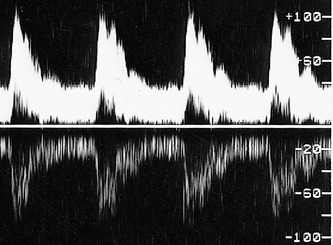
The shape of the Doppler spectrum can also be altered if the high-pass filter is set too high, removing important information from the spectrum, such as the presence of low-velocity diastolic flow. The gain used to amplify the Doppler signal may also alter the appearance of the spectrum. If the gain is set too low, flow may not be detected. Increasing the gain can increase the appearance of spectral broadening, as shown in Figure 6.3, and may also lead to errors in velocity measurements. An inappropriately high gain can also lead to overloading of the instrument, causing poor direction discrimination, and this may result in a mirror image of the spectrum appearing in the reverse direction on the display (Fig. 6.4). The gain should be set so that the signal is detected but saturation, i.e., complete whitening of the signal, as seen on the right-hand side of the signal in Figure 6.3A&B, does not occur.
Intrinsic spectral broadening
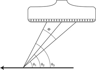
ISB is broadening of the Doppler spectrum that is an artifact, related to the scanner rather than the blood flow interrogated. Linear and curvilinear array transducers use several elements to form the beam (see Ch. 2). Figure 6.5 shows how the ultrasound beam from a linear array transducer can produce a range of angles of insonation, with the Doppler signals being detected at many angles. As the Doppler shift frequency detected is proportional to the cosine of the angle of insonation, θ, this will lead to a range of frequencies being detected even in the presence of a single target. A test object constructed of a string driven at a constant speed by a motor can be used to investigate this effect (Fig. 6.6A). The spectrum obtained from the moving string shows that a large range of Doppler shift frequencies have been detected despite the fact that the target is a single object moving at a constant velocity (Fig. 6.6B). This is due to the range of angles of insonation produced from different elements within the active portion of the probe and is the effect known as ISB. The degree of ISB depends on the range of angles over which back-scattered ultrasound is received by the transducer (Φ in Fig. 6.5) – i.e., it depends on the aperture of the transducer – and on the angle of insonation of the beam (θ).
