Location: Femur (proximal), tibia, craniofacial bones, ribs; then humerus, forearm, pelvis. Same sites in multicentric and polyostotic disease. Frequently more areas in the same long bone, or two to three adjacent bones affected. Lower limb more frequent than upper limb. Hand, foot involved almost only in extensive polyostotic forms. Spine, scapula, clavicle rarely affected. Poliostotic type is usually prevalent in one body side.
Clinical: Monostotic is usually asymptomatic representing an incidental finding. Polyostotic: discontinuous pain (fatigue fractures), bony expansion in superficial bone, pathologic fracture, deformity and lower limb-length discrepancy. In polyostotic forms also cafè au lait spots (“coast of Maine”), multiple endocrine abnormalities (McCune-Albright’s syndrome), intramuscular mixomas (Mazabraud’s syndrome).
Imaging: Standard X-rays show defined defect involving cortical and cancellous areas. Margins well defined, sometimes marked by a rind of bone sclerosis. The cortex is sometimes thinned and expanded but continuous. No periosteal reaction. Radiolucency of “ground glass” appearance is depending upon the amount of intratumoral trabeculae of woven bone. Severe “shepherd’s crook” deformity of the proximal femur, usual in polyostotic form. Isotope scan: rather hot (diffuse dysplastic bone formation) and corresponding to radiographic extent. CT: homogeneity of ground glass radiolucency; cystic cavities and cartilaginous areas (sometimes calcified) when present. MRI: fairly homogenous low signal in T1.
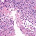
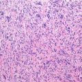
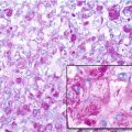
Pathology: Periosteum not involved, underlying cortex is regularly smooth but thin. Lesional tissue, well defined from surrounding bone, whitish to pink, from fibrous to gritty, to hard bony. Sometimes, hemorrhagic areas or cystic spaces with sero-hematic content are present. Rarely, sparse lobules of hyaline cartilage are embedded in the above described tissue. Histology: mixture of benign proliferating fibroblastic cells and islands of woven bone. The bony trabeculae are arranged in a “Chinese letters” fashion. Usually, the bony trabeculae show no clear-cut osteoblastic rimming. Benign giant cells and foam cells are commonly found. No mitotic activity, no atypia. Islands of cartilage may dominate the histologic appearance. May have ABC-like areas.
Course and Staging: Lesions are usually stage 2 in children and adolescents and stage 1 in adults. If a lesion expands and becomes symptomatic in an adult, this may be due to hemorrhage (during pregnancy). Rarely (less than 0.5 % of the reported cases) a sarcoma may develop on a fibrous dysplasia. It usually occurs in adult-advanced age, both in monostotic and polyostotic fibrous dysplasias, more frequent after radiation therapy.
Treatment: Frequently not needed. Curettage and grafting of active lesions should be avoided. Deformities may need corrective osteotomies and internal fixation; preferably by intramedullary devices.
Key Points
Clinical | Incidental findings |
Radiological
Stay updated, free articles. Join our Telegram channel
Full access? Get Clinical Tree
 Get Clinical Tree app for offline access

|


