© Springer-Verlag Berlin Heidelberg 2014
Kyung-Hoi Koo, Michael A. Mont and Lynne C. Jones (eds.)Osteonecrosis10.1007/978-3-642-35767-1_2424. Ficat Staging System
(1)
Department of Orthopaedic Surgery, Seoul National University College of Medicine, Seoul National University Bundang Hospital, 166 Gumi-ro, Seongnam, 463-707, South Korea
24.1 Introduction
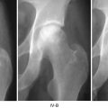
Osteonecrosis of the femoral head is progressive to collapse of the femoral head and secondary osteoarthritis of the hip in 60–80 % of the cases. However, when the necrosis is small and a considerable portion of the weight-bearing dome of the femoral head is viable, the disease is not progressive and patients are doing well for a prolonged time or during their whole life without any medical or surgical intervention. Thus, the classification of osteonecrosis should include the “stage,” which expresses the status of disease progression, and the “extent,” which describes the size of necrosis.
Several different systems to categorize the stage and size of the necrotic portion have been developed and used.
In terms of staging, the Ficat system [1] and ARCO system [2] have been used. For the evaluation of the necrotic extent, the Kerboul method [3] and its modification using MRI [4] and JIC (Japanese Investigation Committee) classification [5] are used.
Steinberg et al. have developed a comprehensive system including both stage and extent [6].
Ficat developed a staging system of femoral head osteonecrosis based on clinical symptoms, functional evaluation of bone, and radiologic findings [1].
Although his staging system was developed in the early 1980s and not based on MRI, it is still in use worldwide because it is simple and easy to use.
24.2 Functional Exploration of Bone
Ficat’s classification was based on two fundamental concepts. The first concept is that all osteonecrosis should pass through a preradiologic stage, which has no specific radiographic appearance. Thus, a radiograph cannot detect early-stage osteonecrosis and a normal radiograph does not necessarily mean a normal hip. Radiographic changes do appear after the reaction of living tissue to the ischemia. The second fundamental concept is that osteonecrosis is the end result of severe and prolonged ischemia. This again presupposes an initial stage in which vascular and medullary abnormality passes undetected by routine radiography.
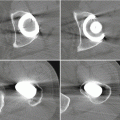
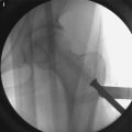
Because MRI was not available in the early 1980s, he emphasized the need for a study of the hemodynamics of the medullary circulation, which he termed the functional exploration of bone (FEB) [7]. Functional exploration of bone is a method for diagnosis of early-stage osteonecrosis. It consisted of three stages: (1) measurement of bone marrow pressure, (2) intramedullary venography, and (3) core biopsy.
Related posts:
 Distinguishing Bone Marrow Edema Syndrome from Osteonecrosis
 The University of Pennsylvania Classification of Osteonecrosis
 Alumina Ceramic-on-Highly Cross-Linked Polyethylene Bearing in Cementless Total Hip Arthroplasty in Young Patients with Osteonecrosis of Femoral Head
 Impaction Bone Grafting
 Causes of Pain in Early and Advanced Stage Osteonecrosis
 Kienböck’s Disease of the Lunate
Stay updated, free articles. Join our Telegram channel

Full access? Get Clinical Tree


