6 Fine-Needle Aspiration Biopsy of Thyroid Nodules
Monzer M. Abu-Yousef
- Palpation-guided fine-needle aspiration biopsy (FNAB) – blind, less accurate; cannot see vessels or cystic versus solid
- Free-hand ultrasound-guided FNAB, without guiding system – cumbersome and less accurate
- Ultrasound-guided core needle biopsy (CNB) using 20-gauge cutting needle
- Ultrasound-guided FNAB with onsite cytopathology – do four aspirations, submit immediately to confirm adequacy
- Ultrasound-guided FNA with syringe using suction – not proven better than capillary aspiration
The purpose of this procedure is to differentiate benign from malignant nodules and to make a specific diagnosis.
- Solid nodules ≥1 cm with questionable ultrasound features
- Nodules with microcalcifications
- Nodules with irregular borders
- Nodules with ill-defined margins
- Solid nodule that is cold on scintigraphy
- Recurrent thyroid mass
- Nodules with cervical lymph nodes suspicious for or harboring thyroid cancer
- Solid nodules ≥1 cm in high-risk patients
- History of neck radiation
- Family history of thyroid neoplasia
- History of multiple endocrine neoplasia (MEN II) syndromes
- Single solid nodule in elderly patient
- Single solid nodule in male patient ≤20 years of age
- Past thyroid lobectomy for contralateral thyroid cancer
- Solid nodule ≥ 1.5 cm
- Complex solid and cystic nodule ≥2 cm
- Nodules showing interval increase in size
- Unsuccessful biopsy of palpable nodule
- Confirm a previously negative thyroid biopsy
- Solid nodule that is cold on scintigraphy
- Multinodular goiter – biopsy up to four nodules with the most suspicious features
- Completely cystic nodules
- Complex nodule with minimal solid component
- Nodules ≤0.5 cm in diameter
- Single solid nodules, hot on scintigraphy
- Nodules with colloid features (comet-tail artifacts)
- Microcystic nodules
- Nodules showing interval decrease in size
- Review patient’s medical records and discuss case with referring physician if necessary
- Review previous imaging studies including ultrasound, positron emission tomography (PET), computed tomography (CT), or magnetic resonance imaging (MRI)
- If thyroid ultrasound exam was not obtained within last 3 months, perform a new one
- No need to check international normalized ratio (INR), prothrombin time (PT), partial thromboplastin time (PTT), or platelet count
- Check if patient is on Coumadin (Bristol-Myers Squibb, New York, NY); if so, check INR
Explain to the patient what procedure will be done, who will be doing it, why it will be done, how it will be done, potential complications, and alternative methods of diagnosis. Answer the patient’s questions and have the patient sign the consent form before signing it yourself.
Current ultrasound machines that are equipped with 6–8 MHz, phased, linear-array transducers are adequate for biopsy purposes.
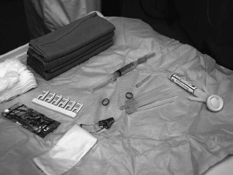
- Place the patient supine with head extended and shoulders supported by a pillow (Fig. 6.2)
