6 First Dorsal Compartment Tendonitis
♦ Setup
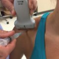
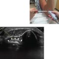
• The patient is seated, facing the operator, with the forearm resting on the table (Fig.6.1).
• The forearm should be in neutral rotation with the thumb up and the ulnar side of the hand on the table.
• A standard shoulder or narrower probe should be used.
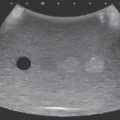
• The ultrasound depth is set at 1.8 cm or less (Video 6.1).

Fig. 6.1 Patient positioning for examination for first dorsal compartment tendonitis.
♦ Landmarks
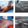
Fig. 6.2 identifies important wrist landmarks, such as Lister’s tubercle and the radial artery. Identification of the radial styloid, scaphoid tubercle, and cephalic vein is also helpful before injection.

Fig. 6.2 Wrist landmarks. APL, Abductor pollicis longus; ECRB, extensor carpi radialis brevis; ECRL, extensor carpi radialis longus; ECU, extensor carpi ulnaris; EDC, extensor digitorum communis; EDM, extensor digiti minimi; EIP, extensor indicis proprius; EPB, extensor pollicis brevis; EPL, extensor pollicis longus; FCR, flexor carpi radialis; FCU, flexor carpi ulnaris; FDP, flexor digitorum profundus; FDS, flexor digitorum superficialis; FPL, flexor pollicis longus. (Reproduced from Janis JE. Essentials of Plastic Surgery. 2nd ed. New York: Thieme Medical; 2014.)
♦ Probe Positioning
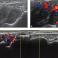
Axial
• The bony landmarks should be palpated and the radial artery identified. The tendons of the first compartment are often palpable at rest. Thumb extension can sometimes help.
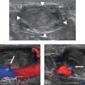
• The probe should be oriented 90 degrees to the long axis of the forearm and parallel to the floor (Fig. 6.3).
• Depending on the curvature of the patient’s wrist and the size of the probe used, the edges of the probe may not contact the skin.
• The probe should be centered over the first dorsal compartment tendons. The operator should scan proximally and distally.
• Color mode is used to identify the nearby cephalic vein and radial artery.
Longitudinal
• Longitudinal positioning is infrequently used.
• The probe should be oriented almost in line with the long axis of the forearm and parallel to the floor.
• The probe is centered over the first dorsal compartment tendons.
• Color mode can be used to identify the cephalic vein.